Case 51
Clinical History, part 1: A 45-year-old man was brought by his wife to the emergency room with a one-week history of nausea, vomiting and a high fever. His wife stated that he had been confused since he awakened that morning. Further questioning revealed that he had a history of alcoholism and had been on a drinking spree two weeks earlier. Physical exam revealed dry mucus membranes, an elevated heart rate of 120 bpm, and orthostasis.
Clinical History, part 2: More detailed examination demonstrated decreased breath sounds and dullness to percussion at the base of the right. Laboratory values were significant for a WBC of 24,000 (60% neutrophils, 24% bands/immature neutrophils, 3% eosinophils, 13% lymphocytes).
Clinical History, part 3: He was admitted to the hospital for observation, further testing, and treatment. However, while being transferred to the in-patient ward, he experienced cardiorespiratory arrest and could not be resuscitated. Gross and microscopic images from the autopsy are provided.
Path Slide 51
[ImageScope] [WebScope]
Image Gallery:
(Summary of Gross and Lab Findings - click here)
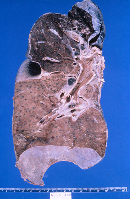
Gram stain of sputum obtained before death shows Gram positive cocci in pairs. The right lung was heavy weighing 700 grams. Its lower lobe showed diffuse gray consolidation. The trachea and bronchi contained a great deal of mucus, and the mucosa was dark red.
|
(Summary of Microscopic Findings - click here)
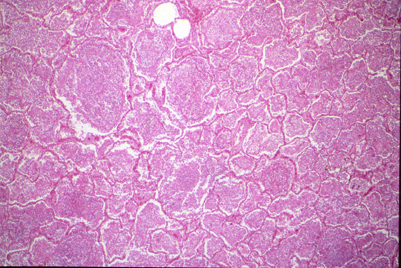
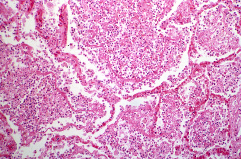
The alveoli are distended and contain a large amount of inflammatory exudate, which consists of many polymorphonuclear leukocytes, a few RBC's, macrophages and strands of fibrin. Many RBC's have been phagocytosed by the macrophages and are undergoing disintegration. The alveolar septa are delicate and well preserved, but markedly congested.
|
(Review Normal Histology - click here)
Norm No. 24 Lung
[ImageScope] [WebScope]
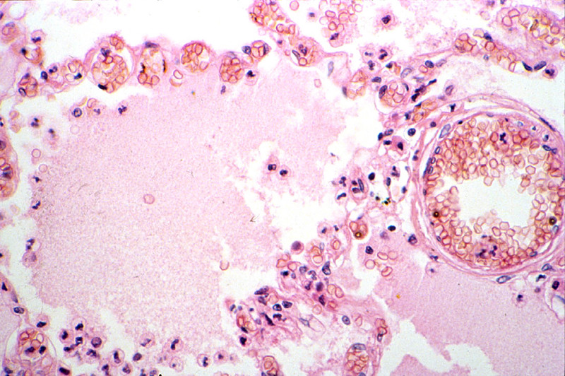
The primary function of the lung is gas exchange. Therefore, alveoli have thin walls lined by thin flat pneumocytes and endothelial cells. There is no thickening or fibrosis of the interstitium. The bronchioli are lined with basally oriented ciliated columnar epithelium. The bronchi are lined by similar epithelium. There are mucous glands within the submucosa. The bronchial smooth muscle is not hypertrophied. The pulmonary vessels are patent with no evidence of intimal thickening or muscular hyperplasia.
|
|
|