CASE NUMBER 300, part 2

Clinical History: A 14-year-old girl is brought to the emergency department with a 1-day history of fevers and decreased consciousness. She had been well until the previous day when she awoke with left-sided headache, anorexia, fatigue, and subjective fevers. During the course of the day, her level of consciousness decreased. In the ED, her temperature is 38.8°C, and she is difficult to arouse. She resists neck flexion, but the remainder of her physical examination is unremarkable.


Clinical History, Part 2: Labs are drawn resulting in the following:

  • CBC: white blood cell count of 29.3×10⁹/L (93% neutrophils and 7% lymphocytes).
  • Lumbar puncture results include
    • a nucleated cell count of 7200×106/L, (92% neutrophils, 7% lymphocytes and 1% monocytes),
    • protein level 1.79 g/L (normal 0.2 g/L to 0.40 g/L)
    • glucose level 2.3 mmol/L (2.2 mmol/L to 3.8 mmol/L).


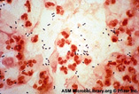
Clinical History, Part 3: A sample of CSF fluid is submitted for Gram staining and blood cultures are drawn. The results of the CSF Gram stain are shown below.

Image Gallery:

Summary of Gram stain and blood culture results

The CSF Gram stain showed Gram-positive diplococci, subsequently growing S. pneumoniae. A peripheral blood culture revealed the same organism.

Diagnostic lab image questions:

  • Describe the Gram stain. How does this affect your differential diagnosis?

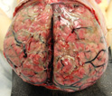
Clinical history (conclusion): Head CT scan shows opacification of the left mastoid with no obvious bony destruction. She is admitted to the pediatric ICU, where she is treated with intravenous vancomycin, ampicillin, and ceftriaxone. Despite aggressive therapy and supportive care, the patient never regains conciousness. She develops intractable seizures and hemodynamic instability and dies three days after hospital admission. Gross and microscopic images from specimens obtained at autopsy are shown.

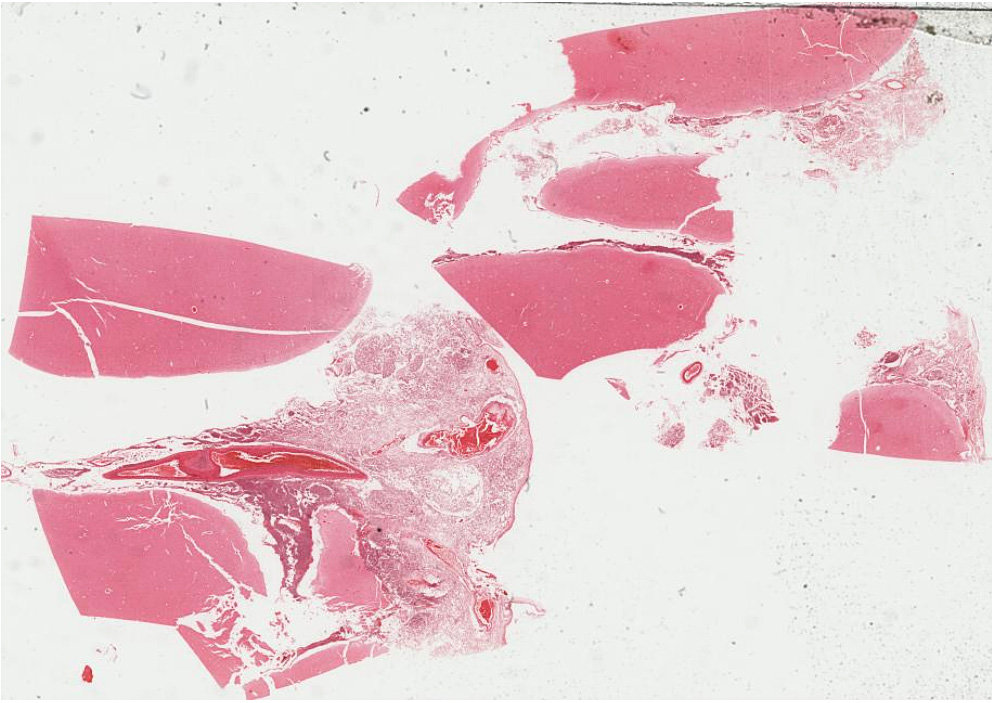
Virtual Slide (slide courtesy of IndianaU): [DigitalScope]

Image Gallery:

Summary of Gross Findings

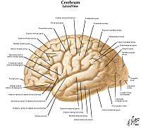
There is thickening and opacification of the meninges. Pus can be seen filling the sulci.

Summary of Microscopic Findings
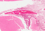
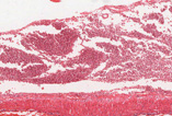
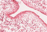
A neutrophilic exudate can be seen in the meninges. Neutrophils in the subarachnoid space can infiltrate and damage cranial nerves leading to focal deficits. Invasion into leptomeningeal blood vessels may lead to thrombosis and ischemic infarction. Blood vessels are dilated and congested. If the infection resolves, the fibrinopurulent exudate can organize into fibrous tissue that blocks the exits of the fourth ventricle and impairs CSF flow around the cerebral convexities, which may lead to hydrocephalus. Bacteria and neutrophils don’t usually gain access to the brain parenchyma due to the glia limitans, a thick mesh of astrocytic processes joined by dense junctions; therefore, brain abscesses are only a rare complication.
(Review Cerebral Cortex Histology)
Cerebrum
Slide 76
(cerebrum, luxol blue/cresyl violet) [DigitalScope]
Slide 76b (toluidine blue & eosin) [DigitalScope]
  
The cerebral cortex is loosely stratified into layers containing scattered nuclei of both neurons and glial cells.  Examine the layered organization of the cerebral cortex using slide 76 stained with luxol blue/cresyl violet [ORIENTATION] (which stains white matter tracts and cell bodies) or toluidine blue and eosin [ORIENTATION] (TB&E, toluidine blue stains the nuclei and RER of cells whereas eosin stains membranes and axon tracts).  Typically one or more sulci (infoldings) will extend inward from one edge of the section.  Examine the gray matter on each side of the sulcus using first low and then high power.  Neurons of the cerebral cortex are of varying shapes and sizes, but the most obvious are pyramidal cells.  As the name implies, the cell body is shaped somewhat like a pyramid, with a large, branching dendrite extending from the apex of the pyramid toward the cortical surface, and with an axon extending downward from the base of the pyramid.  In addition to pyramidal cells, other nuclei seen in these sections may belong to other neurons or to glial cells also present in the cortex.  You may be able to see subtle differences in the distribution of cell types in rather loosely demarcated layers. There are 6 classically recognized layers of the cortex:

  1. Outer plexiform (molecular) layer: sparse neurons and glia
  2. Outer granular layer: small pyramidal and stellate neurons
  3. Outer pyramidal layer: moderate sized pyramidal neurons (should be able to see these in either luxol blue or TB&E-stained sections)
  4. Inner granular layer: densely packed stellate neurons (usually the numerous processes aren’t visible, but there are lots of nuclei reflecting the cell density)
  5. Ganglionic or inner pyramidal layer: large pyramidal neurons (should be able to see these in either luxol blue or TB&E-stained sections)
  6. Multiform cell layer: mixture of small pyramidal and stellate neurons

Pyramidal cells in layers III and V tend to be larger because their axons contribute to efferent projections that extend to other regions of the CNS –pyramidal neurons in layer V of motor cortices send projections all the way down to motor neurons in the spinal cord!

Deep to the gray matter of the cerebral cortex is the white matter that conveys myelinated fibers between different parts of the cortex and other regions of the CNS. Be sure you identify the white matter in both luxol blue and TB&E-stained sections, as it will appear differently in these two stains. Review the organization of gray and white matter in cerebral cortex vs. spinal cord.

Gross image questions:

  • Describe the appearance of the brain at autopsy. How does this affect your differential diagnosis?

VM image questions:

  • Identify an area of the brain on the slide that is consistent with the clinical history and gross findings. Take a screenshot and describe what you have identified.