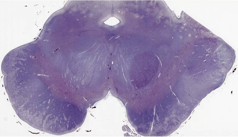
CASE NUMBER 543 - slide courtesy of UMich, PhosphoTungstic Acid-Hematoxylin (PTAH) stain
[DigitalScope]
--Click here to view a video debrief regarding the pathology findings presented in this case--
Clinical History: A previously healthy 62-year-old right handed man noticed that his right hand trembles while relaxed. His wife has also noted that his handwriting has become smaller over the course of the past year. He has no memory deficits, and although hypertensive he never had a stroke.
He does feel fatigued, but can still accomplish his activities of daily living. He has mild difficulty getting out of a taxi. His wife noticed reduced right arm swing with walking, but no falls. They have not noticed any cognitive decline. His wife mentioned he had been having “nightmares” for several years in which he would seem to act out dreams while asleep.
Medications: Lisinopril (for blood pressure). No known allergies
Social: denies use of alcohol, tobacco, or illicit drugs
There is no family history of neurologic problems.
Physical exam:
Vital signs normal
General exam unremarkable
Mental status, cranial nerves, motor, reflexes, sensory, and cerebellar function was normal, with the following exceptions:
Decreased facial expression and reduced blink frequency. Voice soft and monotone.
Intermittent right-sided rest tremor of the arm without postural or kinetic components. Increased tone in both arms, worse on the right. Cogwheel rigidity was detected bilaterally, more pronounced on the right. Bilateral bradykinesia was found with finger tapping and pronation-supination of the hands, more prominent on the right. He walked with stooped posture, smaller shuffling steps, and reduced arm swing more notable on the right. He took 5 steps to turn around. Postural reflex testing was normal.
Tesing and Diagnosis:
Testing
Labs: CBC, LFT’s, BMP, Thyroid function are normal
Imaging: brain MRI is normal
Clinical diagnosis of Parkinson’s disease based on presence of 3 cardinal features:
Tremor
Bradykinesia
Rigidity
Clinical Presentation:
Cardinal Features:
Resting tremor
Bradykinesia
Rigidity
Symptoms that can precede the onset of PD symptoms:
Sleep dysfunction (REM behavior disorder in particular)
Olfactory dysfunction
Constipation
Other Features:
Postural instability
Shuffling gait with reduced arm swing
Stooped posture
Hypomimia
Hypophonia
Dysphagia
Micrographia
Autonomic disturbances: hypotension, constipation, gastroparesis, erectile dysfunction
Cognitive impairment/dementia
Case conclusion: The patient dies in an automobile accident. Gross and microscopic specimens are obtained at autopsy.
Differential diagnosis and pathogenesis:
543-1. What is the differential diagnosis?
543-2. What is the pathogenesis of this disease?
Image Gallery:
Review Midbrain Histology
Midbrain
Slide 42 (midbrain, H&E/luxol fast blue) [DigitalScope]
This slide is an axial section showing one half of the midbrain at the level of the exit of CN III (oculomotor nerve), seen in the upper left of the slide --the orientation of the slide is such that the midbrain turned sideways with the dorsal aspect of the midbrain to the right and ventral is to the left. As such, the superior coliculus is to the right and the cerebral peduncle is to the left. The cerbral aqueduct, lined by ependymal cells, is seen in the upper right portion of the specimen. Just ventral (left) to the cerebral aqueduct is a collection of large neurons corresponding to the oculomotor and Edinger-Westphal nuclei. Ventral and lateral to those nuclei is the white matter tract of the medial lemniscus. Ventral to the medial lemniscus are fiber tracts associated with the superior cerebellar peduncle and portions of the red nucleus. Sandwiched between the cerebral peduncle and the superior cerebellar peducle/red nucleus is the SUBSTANTIA NIGRA, which contains prominent, pigmented neurons corresponding to the pars compacta.
|
|
Summary of Gross Findings
An axial section through the midbrain illustrates pallor of the substantia nigra. The midbrain of the patient is displayed on the right. A normal midbrain is shown on the left.
|
Summary of Microscopic Findings
This is a phosphotungstic acid stain of the midbrain that shows loss of pigmented (catecholeminergic) Viable pigmented neurons contain eosinophilic cytoplasmic inclusions with a halo (Lewy bodies)
|
Gross image questions:
- How does the gross section of the patient’s midbrain differ from a healthy midbrain?
VM image questions:
- On an annotated screenshot, identify pigmented catecholaminergic neurons and a Lewy body.
|

