|
|
Hematologic system Slide list:
Slide Descriptions Specimen No. 74. Peripheral blood smear, AML, human, Wright-Giemsa This specimen of peripheral blood is NOT normal. It was obtained from a patient with a hematologic malignancy (acute promyelocytic leukemia) that resulted in the circulation of immature cells that are normally present only in the bone marrow. The purpose of this slide is to give you some experience in distinguishing these immature white blood cells from the mature white blood cells usually seen in the blood.
Specimen No. 75. Peripheral blood smear, CLL, human, Wright-Giemsa Again, this blood smear is NOT normal. It is from a patient with a hematologic malignancy (lymphoma) that resulted in approximately 50% circulating eosinophils. Since eosinophils are usually rare in peripheral blood, examination of this slide provides you a chance to better learn to recognize these cells.
Specimen No. 82. Peripheral blood smear, normal, human, Wright-Giemsa This slide demonstrates the normal appearance and cellularity of a peripheral blood smear.
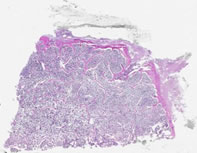
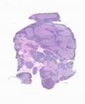
Specimen No. 67. Bone marrow, human, H&E A thin layer of compact/dense bone borders this section, with internal cancellous/spongy bone. The spaces between bony spicules are filled with hematopoietic cells. Not the large numbers of (round, empty) fat cells present within the marrow space. The fat is lost during processing into paraffin sections. Megakaryocytes can be readily identified, since they are approximately 5-fold larger than most bone marrow cells and contain strongly eosinophilic cytoplasm and multiple nuclei. Can you distinguish the myeloid cells from the erythroid cells in the marrow space?
Specimen No. 40. Thymus, human, H&E Each lobule of the thymus contains a basophilic cortex and a less basophilic medulla. The cortex contains numerous densely packed lymphocytes. The medulla has fewer lymphocytes, which allows the visualization of epithelioreticular supporting cells and macrophages, which can be difficult to distinguish from each other. The medulla also possesses numerous eosinophilic and paucicellular structures called Hassall’s bodies (Hassall’s corpuscles) that are characteristic of the thymus. Hassall’s bodies are composed of terminally differentiated thymic epithelial cells. For another comparison, refer to Slide 58, which also contains some tissue from the thymus.
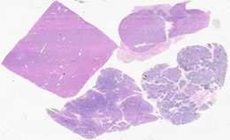
Specimen No. 58. Thymus, pituitary, pancreas, and liver, human, H&E The pancreas is basophilic at low power due to the high content of rough endoplasmic reticulum and Golgi that is necessary for protein production and secretion. The acini are well preserved in this section and the intralobular and interlobular ducts with their simple columnar epithelium are also prominent. The pale eosinophilic areas are the Islets of Langerhans. The thymus is also basophilic at low power due to the high concentration of nuclei (lymphocytes have very little cytoplasm). Lobules contain a cortex of densely arranged thymocytes in the outermost portions, with less densely populated (paler) medulla in the center. Hassall’s bodies are located within the medulla. See also slide notes for Slide 40.
Pituitary: See slide notes for Slide 54 for these sections. This particular piece of tissue shows mostly anterior pituitary.
The square piece of liver is eosinophilic at low power due to the large amount of cytoplasm in the hepatocytes. Note the small, fat globules in some hepatocytes. Identify portal tracts and their component parts (hepatic artery, portal vein, and a bile duct). Note the ray-like pattern of the hepatocytes and the sinusoids that lead to the central vein.
|
||