|
|
Female reproductive system Slide list:
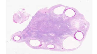
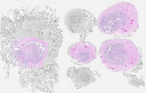
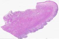
Slide Descriptions Specimen No. 1. Ovary/fallopian tube, human, H&E The surface of the ovary is covered by a single layer of cuboidal epithelium, called the germinal epithelium. Follow the periphery of the ovarian tissue around the section. Small foci of germinal epithelium are visible, however, most of the germinal epithelium is not evident in this section. The germinal epithelium is continuous with the mesothelium that covers the mesovarium (the attachment of the ovary to the mesentery). The dense irregular connective tissue located between the germinal epithelium and the underlying cortex is called the tunica albuginea. Unlike the tunica albuginea of the testis, the ovarian tunica albuginea is not a distinct layer. The ovarian stroma is highly cellular, with a predominately spindle cell morphology. The ovarian medulla is located in the central portion of the ovary (this section is cut somewhat asymmetrically) and consists of loose connective tissue, convoluted blood vessels, lymphatics, and nerves. The endothelium on many of these vessels is well preserved and the smooth muscle layers on many arteries are easy to distinguish. The elastic laminae are not as readily apparent in these vessels, however. Clusters of follicles are seen superficially in the cortex of this section of ovary. Note the small primordial follicles containing a pale oocyte with a prominent nucleolus, surrounded by a single layer of squamous to cuboidal follicular cells. Some sections also contain a primary follicle with several layers of surrounding follicular cells and a prominent thick eosinophilic layer (the zona pellucida) surrounding the oocyte. Multiple secondary follicles are present, many surrounded by hemorrhage. This is a normal occurrence after ovulation; however, the presence of multiple hemorrhagic regions indicates some abnormality. Theca interna and externa cells can be seen at the periphery of some of the secondary follicles. An old corpus luteum (also called a corpora albicans) can also be found. This region is weakly eosinophilic and has a fluffy shape (which will become scarred down with time). A single cross-section of fallopian tube is also located on this slide. Note the ciliated simple columnar epithelium with a complex folding pattern.
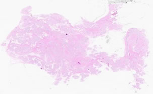
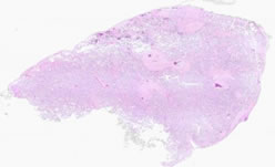
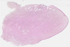
Specimen No. 8. Breast, human, H&E This breast is composed of mainly fibrous connective tissue and a small amount of adipose tissue. Circular collections of glands (acini) are arranged into lobules. Note that the connective tissue between acini within a lobule is more thin and delicate than the dense connective tissue surrounding the lobules. The inactive glands on this slide are composed of two cell thick epithelium (cuboidal with underlying myoepithelium) in the small acini and ducts. The myoepithelium is found between the uppermost epithelial layer and the basal lamina and is the basis for the contractile nature of the duct. A two-layered epithelium may be more easily discerned in the larger ducts. The predominance of fibrous tissue and the small size of the lobular units are indicative of a small breast from a non-pregnant/non-lactating woman. Larger breasts contain more adipose tissue. Compare this to Slide 59, showing lactational change.
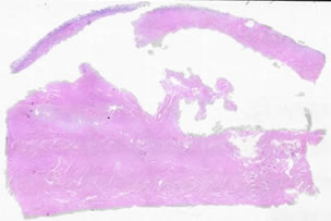
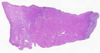
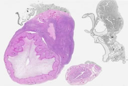
Specimen No. 19. Uterus, human, H&E The majority of the tissue seen on this slide is myometrium, which is composed of bundles of smooth muscle. Spiral endometrial glands cut both longitudinally and in cross-section are evident at the surface, which has become detached from the rest of the uterine wall. Note the prominence of nerves and vessels amid the interlacing bundles of smooth muscle.
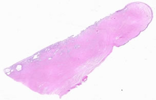
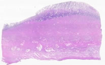
Specimen No. 20. Cervix, human, H&E Focus on the surface epithelium of the cervix. At the uterine end of the cervix (the endocervix), the epithelium is simple columnar, whereas a stratified squamous epithelium is present in the exocervix closer to the vagina. Note the squamo-columnar junction; the hemorrhage present is not normal, nor are the other areas of inflammation. Note the lack of keratinization of this stratified squamous epithelium, which helps to differentiate it it from the epidermis of skin. Cervical glands, which secrete a mucous substance to block entrance to the uterus or to facilitate the entry of sperm depending on the stage of the menstrual cycle and to lubricate the vagina, can be seen in this slide.
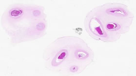
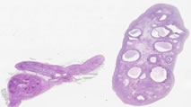
Specimen No. 21. Fallopian tube, human, H&E Six cross-sections of a fallopian tube can be observed in this slide, although all cannot be viewed at the higher magnifications. At low magnifications, the intricate folding of the mucosa (ciliated columnar epithelium) is noticeable in all sections. However, the extensiveness of the folds increases as the sample is taken from further away from the uterus. Deep to the mucosa is a thick layer of circular smooth muscle, a thin layer of longitudinal smooth muscle, as well as a highly cellular connective tissue and vasculature. Note that there is no debris in the lumen of the oviduct as there is in the seminal vesicles.
Specimen No. 29. Chorioamnionic membrane, human, H&E The membranes have been rolled and cut in cross-section to provide more area for evaluation. Examine the membrane roll on the right at higher magnification. Note the simple cuboidal epithelial lining of the amnion. The chorion is the more cellular, vacuolated layer deep to the amnion that carries the fetal vasculature.
Specimen No. 30. Placenta, human, H&E Note the chorionic villi, cut in cross-section, with their 2 layer covering of outer syncytiotrophoblast and inner layer of cytotrophoblast (the cytotrophoblast layer may only be evident focally). Clusters of syncytiotrophoblast cells called syncytial knots are also present. Branches of the umbilical arteries and veins are located within the chorionic villi. The deeply eosinophilic acellular material present on the surface of some villi is fibrin. Calcification, seen as bluish crystalline deposits, is present focally and is one indication of a mature placenta.
Specimen No. 31. Umbilical cord, human, H&E Note the vessels that are present: two arteries and one vein. The two arteries carry blood away from the fetal heart to the placenta and the vein is involved in returning blood to the fetus. The umbilical arteries lack an internal elastic lamina, and have a double layered muscular wall. An internal elastic lamina can be seen in the vein, which typically has a larger diameter than the two arteries. The muscle in the vein is thinner than in the artery, and consists of a single layer of circular smooth muscle. The connective tissue of the umbilical cord consists of Wharton's jelly which protects the umbilical vessels. Wharton's jelly is primarily hyaluronic acid and chondroitin sulfate, with delicate interlacing microfibrils and sparse collagen. The surface of the umbilical cord is a single layer of cuboidal to squamous amnionic epithelium.
Specimen No. 52. Cervix, human, H&E The cervix essentially bridges the uterus and the vagina, and thus changes in the histology of this tissue can be observed along its length. The mucosa of the external os (exocervix) contains stratified squamous epithelium that is similar to the vagina. The internal os (endocervix) has simple columnar epithelium that is similar to the lining of the uterus. Note that glands are only apparent underlying the endocervical mucosa. Furthermore, compare the tissue that underlies the epithelial surfaces in both regions. What differences do you observe?
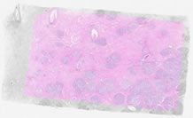
Specimen No. 59. Breast with lactational change, human, H&E Compare this slide with that of normal breast seen in Slide 8. The acini in this slide are convoluted and markedly enlarged relative to non-lactating breast. Lobules likewise seem to contain more acini than in non-lactating breast. Note the eosinophilic secretions in the lumen. The background stroma in this tissue is predominantly collagenous fibrous tissue, rather than the adipose tissue which is more commonly seen in breast biopsies.
Specimen No. 76. Uterus/endometrium, human, H&E The endometrium on this well-oriented section of uterus shows tall columnar surface epithelium, with spiral glands extending down into the highly cellular endometrial stroma. The underlying myometrium consists predominately of bundles of smooth muscle, with prominent blood vessels and nerves. Note the presence of more connective tissue near the serosal surface.
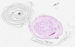
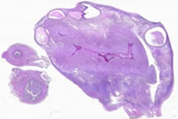
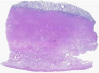
Specimen No. 77. Ovary/fallopian tube, human, H&E The most prominent structure in this ovary is the corpus luteum. The corpus luteum is composed of oval to polygonal, highly eosinophilic cells with abundant cytoplasm and prominent nucleoli. The ovarian medulla containing many blood vessels is also prominent in this section. Two sections of fallopian tube are present. Note the vessels, nerves, and smooth muscle present in the wall of the fallopian tube. One of the tubes also shows a small paratubal fluid-filled cyst. Examine the epithelium using the high dry or oil objective to visualize the cilia present on the columnar epithelial cells.
Specimen No. 78. Cervix, human, H&E Follow the epithelial surface of this tissue around the entire section. Note the transition from the stratified squamous epithelium of the exocervix to the columnar epithelium of the endocervix at the squamo-columnar junction. The endocervix has abundant underlying glands that provide cervical mucus. Identify the smooth muscle present deeper in the section.
Specimen No. 79. Uterus/endometrium, human, H&E This section is cut tangentially, causing the endometrial thickness to appear much thicker at one end of the section. Some slides also show an artifactual lack of staining in the upper endometrium. This endometrium is secretory, day 20-22.
Specimen No. 80. Uterus/endometrium, human, H&E The secretory endometrial glands profiles are complex. Spiral arteries are prominent.
Specimen No. 81. Ovary/adrenal, human, H&E The ovary contains numerous follicles in various stages of maturation. Primordial follicles, consisting of an oocyte surrounded by a single layer of squamous epithelium, are numerous in the cortex. Some early primary follicles can also be seen. Most of the prominent follicles are the secondary follicles. Although the oocyte may not be present in all sections, one mature follicle with a well-developed cumulus oophorus can be identified.
Specimen No. 83. Ovary with corpus luteum/fallopian tube, human, H&E This is an excellent example of an ovary with a prominent corpus luteum occupying approximately 50% of the organ. Two sections of fallopian tube are also present on this slide. Can you identify the paratubal cysts? Although not strictly normal, these tubal cysts are prevalent and rarely cause problems for the patient.
|
||
Click here to submit questions or comments about this site. Updated 10/28/14 - Velkey |
||