|
|
Lymphoid system Slide list:
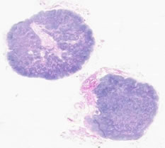
Slide Descriptions Specimen No. 9. Lymph nodes, human, H&E Two lymph nodes are present on this slide. Dense connective tissue forms the capsule that surrounds the lymph node. Trabeculae are contiguous portions of the capsule that penetrate into the cortex. The subcapsular sinus is immediately below the capsule and, can be seen as a space between the capsule and the lymph node parenchyma. This slide is NOT a wonderful example of classic germinal centers (see Slide 18 instead); however, the hilum with its blood vessels and lymphatics is quite obvious. Notice the dramatic differences between the architecture of the cortex and the medulla. The sinuses in these lymph nodes are filled with histiocytes which have pale eosiniphilic cytoplasm, making these regions stand out as pale regions in the lymph node medulla. See also Slide 22. Take a close look at blood vessels present within the lymph node. Many post-capillary venules with high (almost cuboidal) endothelium are present. These vessels are specialized to provided lymphocyte egress from the blood into the lymph node. For another comparison, refer to Slide 57, which contains some lymph node tissue.
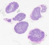
Specimen No. 18. Lymph node, human, H&E The major difference between this slide and Slide 9 is the presence of prominent germinal centers in these lymph nodes. See also Slide 22. For another comparison, refer to Slide 57, which contains some lymph node tissue.
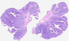
Specimen No. 32. Spleen, human, H&E The dense capsule surrounding the spleen can be seen under low power; however, trabeculae are not readily apparent in this sample. Despite this downfall, the slide clearly illustrates the differences between white pulp (collections of lymphocytes) and red pulp where red blood cells are filtered. Furthermore, a thin ring of lymphocytes around central arteries can be seen in several fields of view; these are referred to as periarteriolar lymphatic sheaths. For another comparison, refer to Slide 57, which contains some tissue from the spleen.
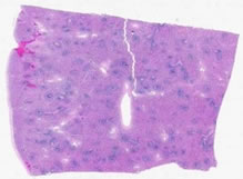
Specimen No. 34. Tonsil/salivary gland/skeletal muscle, human, H&E The stratified squamous epithelium that covers the tonsil frequently dips into the underlying connective tissue, forming tonsillar crypts. The lymphoid cells within the tonsil can be seen either in aggregates or arranged as nodules containing germinal centers. A mucous-secreting salivary gland and a piece of skeletal muscle can also be seen in this section. Where is the muscle from?
Specimen No. 57. Adrenal, spleen, lymph node and thyroid, human, H&E Refer to the slide notes for slides 7, 9, 18, 22, and 32 for descriptions of adrenal, spleen and lymph node (copied below). The densely packed follicles that comprise the bulk of the thyroid are most notable for the homogeneous mass of colloid in the center. Note that both brown and white fat are present around the adrenal gland.
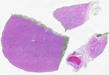
Specimen No. 68. Prostate/nerve ganglia/spleen, human, H&E The largest piece of tissue on this slide is the spleen. The splenic capsule is composed of dense connective tissue, which dips into the parenchyma to form trabeculae. The white pulp appears blue by H&E due to the large numbers of lymphocytes with very little cytoplasm. Many of these lymphocytes can be seen surrounding the central arterioles (the periarteriolar lymphoid sheath). Central arteries are not always visible in the plane of the section. Note the types of cells present in the red pulp versus the white pulp. The prostate demonstrates somewhat complex glands due to folding. The fibromuscular stroma is well-preserved. Note that numerous nerves and nerve ganglia surround the prostate, which has no true capsule.
|
||