|
|
Male reproductive system Slide list:
Slide Descriptions Specimen No. 5. Prostate/seminal vesicle, human, H&E The prostate is composed of bundles of smooth muscle, with sparse prostatic glands present in this section; note the two layer structure of the gland and the predominance of columnar to cuboidal epithelium lining the mucosal layer. Corpora amylacea, or prostatic concretions, are lamellated bodies found in the prostatic alveoli and are easily identifiable by their eosinophilic nature. The highly innervated nature of the prostate is visible due to the presence of several nerve ganglia in this section; observe under high power to visualize individual neurons. The neurons are large cells, with abundant pale eosinophilic cytoplasm and a prominent single nucleolus. Seminal vesicles are also seen in cross-section in this slide. The seminal vesicle mucosa can appear as "villi" due to the extensive folding. Examination using higher magnification reveals a loose connective tissue layer under the pseudostratified epithelium. Golden-brown lipofuschin pigment is prominent in the seminal vesicles, helping to distinguish this gland from the prostate. The prostatic urethra is also present in this section and is identifiable by the transitional epithelium.
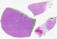
Specimen No. 11. Seminal vesicle, human, H&E Note the pseudostratifed columnar epithelium and the unique appearance of the secretory mucosa that results from the cross-sectioning of extensively folded sacs. The golden-brown lipofuschin pigment characteristic of the seminal vesicle is abundant. The mucosa covers a loose connective tissue layer (lamina propria) which overlays two layers of muscle, one circular and the other longitudinal. Find a nerve ganglion. The black color at the tissue periphery is marking dye that remains from the surgical sample. See also Slide 33.
Specimen No. 33. Seminal vesicle, human, H&E Refer to the notes for Slide 11 (copied below).
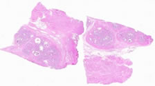
Specimen No. 63. Vas deferens, human, H&E This slide contains two complete cross-sections of vas deferens (a.k.a. ductus deferens). Note the small diameter of these cross-sections by holding the slide up to the light. The vas contains 3 layers: mucosa, muscularis, and adventitia. The mucosa contains pseudostratified columnar epithelium. Stereocilia are present at the mucosal surface, but are not well visualized on this slide. The muscularis is three layered, with inner and outer longitudinal and middle circular layers. The adventitia is composed of loose connective tissue.
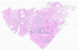
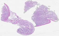
Specimen No. 68. Prostate/nerve ganglia/spleen, human, H&E The largest piece of tissue on this slide is the spleen. The splenic capsule is composed of dense connective tissue, which dips into the parenchyma to form trabeculae. The white pulp appears blue by H&E due to the large numbers of lymphocytes with very little cytoplasm. Many of these lymphocytes can be seen surrounding the central arterioles (the periarteriolar lymphoid sheath). Central arteries are not always visible in the plane of the section. Note the types of cells present in the red pulp versus the white pulp. The prostate demonstrates somewhat complex glands due to folding. The fibromuscular stroma is well-preserved. Note that numerous nerves and nerve ganglia surround the prostate, which has no true capsule.
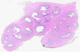
Specimen No. 72. Testis/epididymis, human, H&E At least two pieces of testis are present on this slide. Note the thick capsule, the tunica albuginea. The parenchyma of the testis is composed of seminiferous tubules. Rare sperm are present in the lumen in this 14 year old patient. The epididymis is cut in multiple cross-sections, which demonstrate the pseudostratified columnar epithelium. The long stereocilia are evident, with a prominent terminal bar. These structures are not true cilia and do not "beat".
|
||
Click here to submit questions or comments about this site. Updated 10/24/12 - Velkey |
||