|
|
Nervous system Slide list:
Slide Descriptions Specimen No. 41. Dorsal root ganglion, human, H&E LFB With this modified H&E/Luxol fast blue staining protocol, myelin stains blue and lipofuschin stains yellow. Note the elongated nuclei of the Schwann cells and the satellite cells that surround the neurons. The nucleoli are very apparent within the pale staining nuclei of the sensory neurons.
Specimen No. 42. Midbrain, human, H&E/LFB Features of this section include the 3d cranial nerve and the myelinated cerebral peduncle. Can you find the pigmented neurons of the substantia nigra? These neurons contain neuromelanin, which is a precursor of dopamine. The cerebral aqueduct on this section appears as a cystic space lined by ciliated cuboidal epithelium (the cilia are difficult to see in most sections). You can also see the myelinated fibers of the corticospinal tract, surrounded by the oligodendroglial cells that make the myelin.
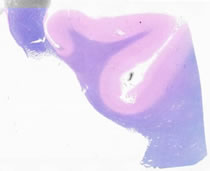
Specimen No. 43. Thalamus, human, H&E/LFB The most important feature of this section is the pineal gland, which contains neurosecretory elements. The mineralization seen is common. In birds, the magnetite (iron) content of the pineal is thought to be important in directing migration. The back of the 3d ventricle, surrounded by the choroid plexus (cuboidal epithelium) is also seen in the lower right of this section.
Specimen No. 44. Cerebral cortex, human, H&E/LFB The arachnoid layer of delicate connective tissue can be visualized focally, along with its underlying pia mater, a single cell layer directly on the cortex. The cerebral gray matter appears pink in this stain. The neurons in the gray matter have a prominent single nucleolus, with basophilic areas of cytoplasm. This is the Nissl substance, which actually is RNA. The white matter appears blue due to its high myelin content. Astrocytes are large and round. Oligodendroglia have a lymphocyte-like appearance.
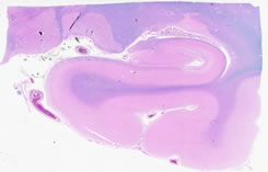
Specimen No. 45. Hippocampus, human, H&E/LFB A branch of the posterior cerebral artery is present in the center of the section. Note also the choroid plexus, consisting of ependymal cells and cuboidal cells. Capillaries are visible in the central parts of the loops. Mineralization of the choroid plexus is commonly observed. The choroid plexus makes the cerebrospinal fluid. Note the large pyramidal neurons of the hippocampus itself. Dentate neurons are arranged in rows. Note that the internal elastic lamina of arteries stains blue with this stain. The small ovoid /spindle cells are microglia; the perineuronal oligodendroglia have nuclei that resemble lymphocytes.
Specimen No. 46. Thalamus, human, H&E/LFB Note the ependymal surface on the medial edge. Six to 12 clusters of neurons (called nuclei) can be seen in this section. The internal capsule is visualized here by its high myelin (stains bright blue with LFB stain) content.
Specimen No. 47. Cingulate gyrus/ corpus callosum, human, H&E/LFB The meninges (arachnoid and pia) can be seen on one side of the tissue, with the ependyma on the other side. The cingulate gyrus has a similar histologic appearance to the cortex.
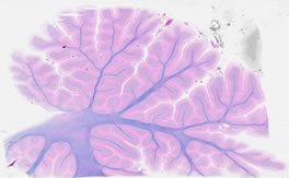
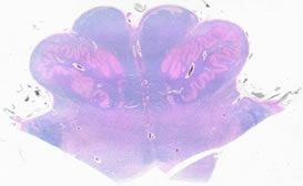
Specimen No. 48. Cerebellum, human, H&E/LFB Note the characteristic folia and gyrae of this tissue. The arachnoid envelopes the folia. The layers are: the relatively acellular molecular layer, the Purkinje cell layer, and the granular cell layer. This is a good place to identify microglia. Myelinated tracts and the ependyma of the 4th ventricle are present on this section.
Specimen No. 49. Medulla, human, H&E/LFB This section demonstrates the floor of the 4th ventricle, pyramidal tracts, the (irregularly shaped) inferior olivary nucleus. Cranial nerve nuclei are visible in the floor of the 4th ventricle.
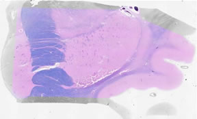
Specimen No. 50. Basal ganglia, human, H&E/LFB The insula resembles cortex histologically. Myelinated tracts include the extreme capsule, the claustrum, and the external capsule. The putamen has a striated appearance. The globus pallidus appears somewhat speckled here. The internal capsule is the very large myelinated tract. The caudate nucleus is also present, with ependyma adjacent.
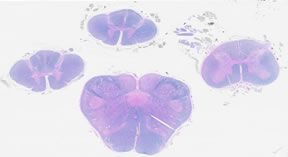
Specimen No. 51. Spinal cord, human, H&E/LFB The 4 sections on this slide are taken from the lower medulla, upper cervical, mid-cervical, and lumbar regions of the spinal cord. Note that both neurons and myelinated tracts are present in these sections. The neurons are present in the gray matter in the central portion of the cord, surrounded by the myelinated axons of the white matter. The roots of various spinal nerves are seen in this section, but not the dura.
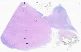
Specimen No. 53. Myenteric plexus, human, H&E A thin connective tissue layer is located between layers of the muscularis externa layer of the gastrointestinal tract. Occasional clusters of neurons can be found in this connective tissue. These are part of the myenteric plexus (Auerbach’s plexus). These neurons are important for coordinating the muscular movements that propel food through the digestive tract (peristalsis).
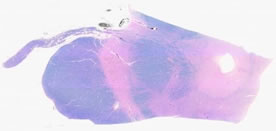
Specimen No. 54. Pituitary, human, H&E and LFB The pituitary gland is differentiated into two regions: the adenohypophysis (anterior) and the neurohypophysis (posterior). The adenohypophysis contains cells that are chromophobes (stain poorly) and chromophils (stain well with either basic or acidic dyes, the basophils and acidophils, respectively). The neurohypophysis is less cellular and contains nonmyelinated nerve fibers and pituicytes (which are comparable to neuroglial cells of the CNS). Some colloid areas stain a brilliant blue. For another comparison, refer to Slide 58, which contains some tissue from the thymus.
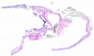
Specimen No. 55. Dural sinus, human, H&E The dural sinuses are simply veins that have an atypical structure due to their location. What is seen in this sample are spaces in the dura mater that are lined with endothelial cells in order to form a venous channel.
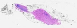
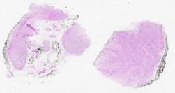
Specimen No. 56. Sympathetic ganglion, human, H&E The most pronounced feature of this slide is the sheer number of neurons that can be observed. What characteristics of this ganglion can you use to differentiate this from ovary? Disregard the marking pigment that can be seen in several fields of view.
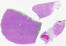
Specimen No. 68. Prostate/nerve ganglia/spleen, human, H&E The largest piece of tissue on this slide is the spleen. The splenic capsule is composed of dense connective tissue, which dips into the parenchyma to form trabeculae. The white pulp appears blue by H&E due to the large numbers of lymphocytes with very little cytoplasm. Many of these lymphocytes can be seen surrounding the central arterioles (the periarteriolar lymphoid sheath). Central arteries are not always visible in the plane of the section. Note the types of cells present in the red pulp versus the white pulp. The prostate demonstrates somewhat complex glands due to folding. The fibromuscular stroma is well-preserved. Note that numerous nerves and nerve ganglia surround the prostate, which has no true capsule.
|
||