|
|
Urinary system Slide list:
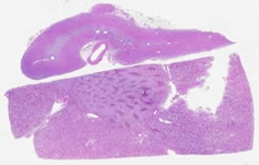
Slide Descriptions Specimen No. 6. Kidney/adrenal, human, H&E The large rectangular piece of tissue is the kidney. Clearly differentiated in this slide is the cortex and medulla of the kidney. The cortex is subdivided into two regions, the cortical labyrinth containing glomeruli (renal corpuscles) and the medullary rays comprised of the straight tubules of the ascending and descending limbs of Henle's loop as well as the collecting tubules. The medulla is composed of the collecting tubules (pale, cuboidal epithelium), and the vasa recta (capillaries that help to maintain osmotic gradients) forming a pyramidal structure. Find the renal pelvis. What type of epithelium lines the lumen? In the separate section of adrenal gland, one can clearly discern the cortex from the medulla. There are three zones in the cortex (zona glomerulosa, zona fasciculata, and zona reticularis); can you distinguish them? Note the superficial resemblance of the structures in the zona glomerulosa to the glomeruli in the kidney.
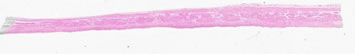
Specimen No. 14. Ureter, human, H&E Notice that under low power, the entire wall of the ureter, as well as the lumen, are in the field of view (therefore this is a tube with a small diameter in vivo). The mucosa is composed of transitional epithelium that is characterized by the dome-shaped cells at the surface. These cells elongate when stretched, providing the ureter with its distensibility. The muscularis of the ureter has three layers: an inner longitudinal layer, a middle circular layer, and an outer longitudinal layer. The outermost layer of muscle can only be seen at the lower end of the ureter. Can you visualize it in this slide?
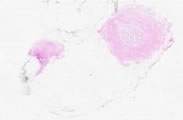
Specimen No. 69. Kidney/adrenal, human, H&E Both cortex and medulla are represented in the adrenal section. The kidney has both cortex, with glomeruli and tubules, and medulla. The medullary rays are present in both longitudinal and cross-section in the central portion of the kidney section. Note the thin fibrous capsule.
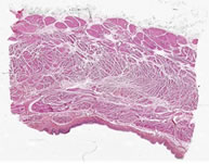
Specimen No. 95. Urinary bladder, H&E The transitional epithelium is located at the bottom of the section. The epithelial layer appears to be 2-5 cells thick, with no particular shape to the upper cells. The lack of the classic “dome shape” of the transitional epithelial cells combined with the relatively thin layers of smooth muscle layer indicate that this bladder was fixed when distended. The lamina propria that underlies the epithelium is composed of dense irregular connective tissue. There is no muscularis mucosae or submucosa. The smooth muscle (the muscularis propria) is arranged in 3 relatively indistinct bundles, oriented at right angles to each other. This is best seen toward the middle of the section, since the uppermost layer (relative to the epithelium that is at the bottom of the section) appears to be cut tangentially over much of the section. Note the different appearance of smooth muscle cut in cross-section vs. longitudinal section. A serosa is present underlying the muscularis propria. The serosa consists of loose fibroadipose tissue that is bounded by a layer of mesothelium arranged in a simple squamous arrangement. The presence of a serosal surface indicates that this section was taken from the superior surface of the bladder that forms part of the base of the peritoneal cavity. See also Slide 97 and Slide 98, which contain urinary bladder tissue.
Specimen No. 97. Urinary bladder, human, H&E Again, the transitional epithelium is located at the bottom of the section. The epithelium has multiple layers and the outer layer contains many dome-shaped cells. This allows identification as transitional epithelium, fixed when the bladder was relatively empty. The layers present are similar to what was described for Slide 95, except that the outermost layer (seen at the top of the section) is irregular and is not bounded by mesothelium. This layer is most correctly referred to as an adventitia and is characteristic of the inferior and posterior surfaces of the bladder that are attached to other pelvic organs.
Specimen No. 98. Urinary bladder, human, H&E The features of this slide are very similar to those shown in Slide 95, including fixation in distension and the presence of a serosa.
|
||