| Normal Lab Values | ||||||
|
|
Week 7: Environmental Pathology |
||
| Suggested readings from Robbins 8th ed. |
|
|
|
|
Pathology Cases for Week 7
Pathology Case Descriptions CASE NUMBER 92 Gross: The lungs were very large and upon palpation there was marked crepitation. Microscopic: The alveolar spaces are distended and the alveolar septa are thickened by fibrous tissue. Numerous "free floating" alveolar septa are present, recognizable as pieces at alveolar wall not connected at either end to adjacent septa. Image Gallery: 92-1. What is the most likely diagnosis?
92-2. What factor contributed most to this patient’s disease?
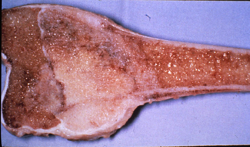
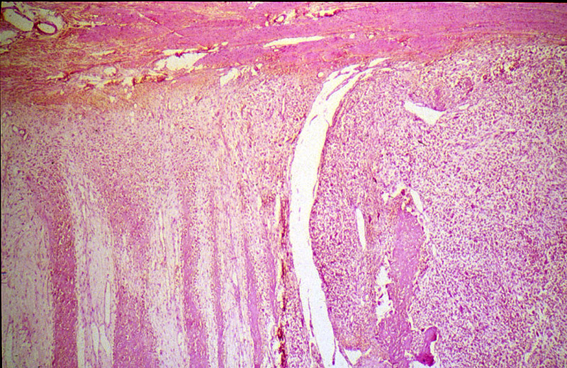
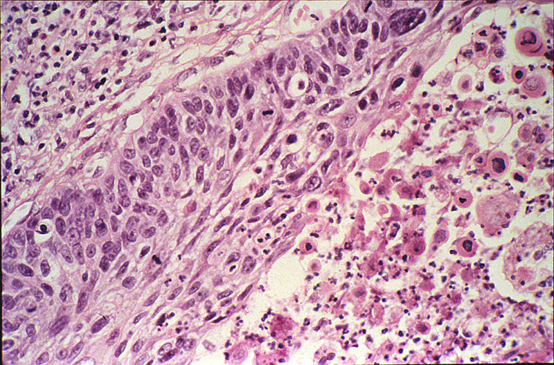
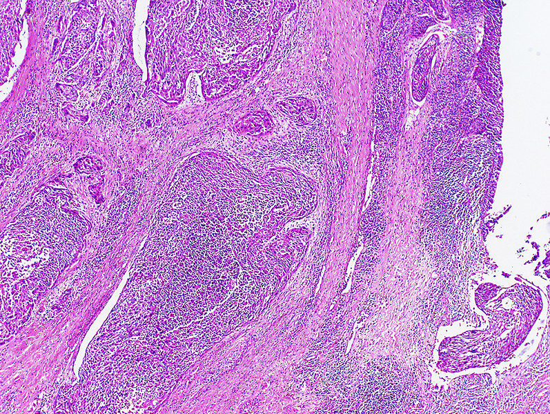
CASE NUMBER 223 Clinical History: A 13 year old male had an enlarging mass in the left thigh for 1 1/2 months. It was not associated with pain or tenderness. The X-ray revealed a large tumor extending around the entire shaft and lower 1/3 or the left femur. Amputation was performed after biopsy. Limb salvage surgery is commonly performed today. Gross: The tumor was a large, pink-gray, soft and granular mass, 8x4x5 cm in size, involving the lower femoral shaft, and growing into the muscle. Microscopic: The tumor is composed of sheaths of loosely packed fibroblastic and osteoblastic cells, which have large hyperchromatic nuclei and scanty eosinophilic cytoplasm. In the intercellular spaces of the tumor there is deposition of pink homogeneous osteoid tissue which frequently transforms into irregular, bony trabeculae. New periosteal bone formation may be seen in the section. Image Gallery: 223-1. This tumor comprises atypical osteoblastic cells that have large hyperchromatic nuclei with nuclear pleomorphism and frequent mitoses, producing new bone matrix (osteoid). The best diagnosis is:
223-2. Mutation and loss of function of the Rb and p53 genes are often encountered in this tumor. Both Rb and p53 are examples of:
223-3. In the assessment of the bone tumor as depicted in these images:
CASE NUMBER 129 Clinical History: A 70 year old white male had an insidious onset of dysphagia of six months duration. X-ray revealed a filling defect and stenosis in the lower esophagus. An esophageal resection was performed. Gross: The resected esophagus contained a firm, raised, pink-gray, friable and necrotic 3.5 x 4.5 cm tumor mass, completely encircling the esophagus and producing a marked stenosis. Microscopic: At one end of the section the squamous epithelium of the esophagus shows marked atypia, pleomorphism and loss of polarity, but no submucosal invasion. These changes represent carcinoma in situ. By following the mucosal lining, one comes upon an area where the cords and nests of poorly differentiated squamous cells are seen to invade the mucosa and submucosa reaching the muscular layer. At one margin of the section the tumor cells are better differentiated with production of keratin and formation of so-called epithelial pearls. Associated with the tumor are marked fibrous proliferation and intense chronic inflammatory infiltration in the stroma. The tumor invades into, but not through the muscularis propria. Image Gallery: What is the most likely diagnosis? 129-1. What is the least important contributory risk factor for this neoplasm?
129-2. The most important risk factor for adenocarcinoma of the esophagus is:
20-3. The condition with the worst 5 year survival is:
CASE NUMBER 154 Clinical History: A 64 year old man who had smoked two packs a day for 40 years, complained of hoarseness and throat pain. A laryngectomy was performed. Gross: An ulcerated tumor involved the left true and false vocal cords and extended across the midline. Microscopic: The mucosa is partially columnar, representing the ventricular mucosa. There is a transition to squamous mucosa with marked nuclear pleomorphism. The abnormal squamous cells extend into the underlying stroma where the cells keratinize. Focal necrosis and an inflammatory infiltrate are present. Image Gallery: 154-1. The histological type of this tumor is best described as:
154-2. Which of the following risk factors is most strongly associated with this disease?
CASE NUMBER 31 Clinical History: This 45 year old man had been well until he was awakened by chest pain that radiated to both arms and neck and was associated with diaphoresis. His blood pressure was 160/110. He was treated with diuretics (Lasix), but he continued to gain weight. Two days after the onset of the chest pain he had a cardiac arrest and died. Gross: The heart was slightly enlarged weighing 460gms. There was severe atherosclerosis of all the major coronary arteries with a recent thrombotic occlusion of the proximal left anterior descending coronary artery. A recent transmural infarct was present in the left ventricle that involved the interventricular septum and the papillary muscle. Microscopic: The slide includes a transmural section of the left ventricle. Nearly the entire section is involved by infarct. However, there is a thin rim (5 to 10 cell layers) of endocardial myocytes which have survived because of diffusion of oxygen and nutrients from the ventricular cavity. Other viable myocytes can be found around larger blood vessels within the section. The intense hypereosinophilia of the necrotic myocytes can best be appreciated by comparing the thin rim of lighter staining subendocardial myocytes with the deeper cells. Note also the karyolysis that is characteristic of coagulation necrosis. In some areas there is little inflammatory response. This observation is explained by microvascular necrosis which does not allow access of circulating leukocytes to these areas. In other areas, especially in the epicardial half of the infarct, there is an intense acute inflammatory response. Many intact neutrophils can be seen. In addition, there are many nuclear fragments from lysed neutrophils. Macrophage activity is not evident. These features of the inflammatory response indicate that the infarct was approximately three to four days old. Note also that the inflammation extends to the epicardial surface and that there are deposits of fibrin on the epicardium. This is called fibrinous pericarditis. The granular grey material seen within some blood vessels is barium sulfate, which was injected to permit post-mortem study of the coronaries by radiography. Image Gallery: What is the most likely diagnosis?
31-1. The event most likely associated with this patient’s problem was:
31-2. What microscopic feature best describes what happened to the nucleus?
31-3. What molecular events led to the increase eosin staining of the dead myocytes?
31-4. The event most likely associated with this patient’s problem was:
31-5. What microscopic feature in this patient indicates that this infarct is at least 24 hours old?
31-6. What microscopic feature is most indicative of reperfusion of ischemic myocardium with irreversible injury?
31-7. What artery was most likely occluded in this patient?
31-8. What myocardium is the most vulnerable to ischemic injury in hypovolemic shock?
31-9. This patient’s myocardial lesion is accompanied by a fibrinous pericarditis, named after:
ENVIRONMENTAL PATHOLOGY Review Items Key Vocabulary Terms (click here to search any additional terms on Stedman's Online Medical Dictionary) LEARNING OBJECTIVES
|
|
Click here to submit questions or comments about this site. Updated 3/12/12 - Velkey |
||