Website assignments:
Additional cases (NOT assigned):
CASE NUMBER 115
[DigitalScope]
Clinical History: A 63-year-old man presented to his primary care physician with a 2-week history of painful lesions on his soft palate that prevent him from eating normally. Physical exam findings are revealed below. A mouthwash was prescribed, but the lesions did not resolve. Three weeks later, additional lesions appeared on the patient's face, chest, and extremities.
115-1. What is the differential diagnosis?
Image Gallery:
Review Skin Histology
Norm No. 15 Skin
[DigitalScope]
Skin consists of keratinizing stratified squamous epithelium. The keratin layer is eosinophilic. Nuclei are not present. Beneath the keratin layer is a layer of flat epithelial cells with small pyknotic nuclei. Cells are regular and not crowded. These keratinocytes rest upon a layer of basal epithelium and a thin basement membrane. Beneath the epidermis is the dermis which consists of loose connective tissue. Within the dermis are sweat and sebaceous glands. Some slides contain hair follicles. There is no inflammation. The blood vessels are patent and do not contain thromboemboli.
|
There are multiple erythematous ulcers of the oral mucosa and round to oval vesicles and flaccid bullae seen on the patients arm.
|
There is a suprabasal blister due to acantholysis (lysis of the intercellular adhesive junctions between neighboring squamous epithelial cells). The basal cells lose their intercellular bridges but they remain attached to the dermis, giving a tombstone appearance. The blister cavity usually contains a few acantholytic cells. The dermal changes are of little significance; there is usually a mild, superficial mixed inflammatory cell infiltrate which usually includes scattered eosinophils.
|
Clinical exam image questions:
- Describe the clinical findings both of the oral mucosa as well as the elsewhere on the patient’s skin. Please use appropriate “dermatologic” terms (e.g., macule, papule, patch, etc.).
VM image questions:
- Describe the histologic findings.
- Based on the image, where was the biopsy taken on the patient? Use a screenshot with annotations to justify your answer.
- At what level in the skin is the blister forming (BP fig 22.8, EP fig 18.15 for reference)? Show in an annotated screenshot an example of acantholysis.
115-2. Antibodies to which of the following are associated with the etiology of this disease?
- BPAG1 and 2
- Desmoglein 3
- Epidermal transglutaminase
- Keratins 14 and 5
- Myelin
115-3. Which of the following is the mechanism of this disease?
- Cytokines and growth factors induce keratinocyte hyperproliferation
- Cytotoxic T cell attack on basal cells
- Cross-reactivity from anti-gluten antibodies
- Delayed type hypersensitivity to urushiol
- Destruction of desmosomes
115-4. Which of the following is characteristic of this disease?
- Acantholysis of superficial epidermis
- Fishnet pattern of IgG in full thickness epidermis
- Granular IgA at the tips of dermal papillae
- Linear pattern of IgG deposition
- Subepidermal blister formation
CASE NUMBER 523 -slide 55938 courtesy of UMich
[DigitalScope]
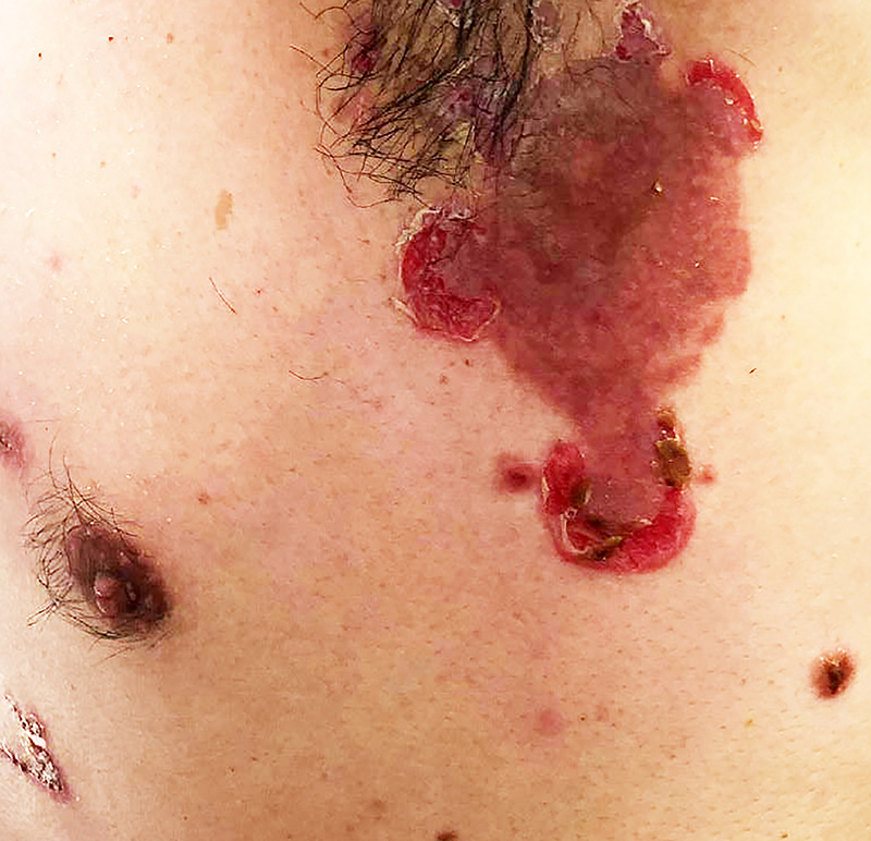
Clinical History: A 45-year-old man presented to his primary care physician with a 2-week history of a bleeding lesion on the back. His wife noticed that the lesion seemed to have been growing over the past several months. The primary care physician suspected a vascular lesion and referred him to the dermatology department for a biopsy.
523-1. What is the differential diagnosis?
Image Gallery:
Review Skin Histology
Norm No. 15 Skin
[DigitalScope]
Skin consists of keratinizing stratified squamous epithelium. The keratin layer is eosinophilic. Nuclei are not present. Beneath the keratin layer is a layer of flat epithelial cells with small pyknotic nuclei. Cells are regular and not crowded. These keratinocytes rest upon a layer of basal epithelium and a thin basement membrane. Beneath the epidermis is the dermis which consists of loose connective tissue. Within the dermis are sweat and sebaceous glands. Some slides contain hair follicles. There is no inflammation. The blood vessels are patent and do not contain thromboemboli.
|
Polypoid growth with variegated colors (black to brown to dark red) and superficial ulcer.
|
The sections show a compound proliferation of atypical melanocytes involving the epidermis and dermis. Ulceration is noted in the epidermis (absence of epidermis with collection of serum and inflammation). The severely atypical melanocytes are arranged as single cells and nests in the epidermis with focal pagetoid spread. The dermis is distended by melanocytes lacking maturation (cells do not decrease in size from papillary to reticular dermis). High power view of the melanocytes show abundant eosinophilic cytoplasm, with high N/C ratio and prominent nucleoli. Mitotic figures are seen.
|
Clinical image questions:
- Describe the clinical findings in the first image. Please use appropriate “dermatologic” terms (e.g., macule, papule, patch, etc.) in your description
- What is your differential diagnosis based on the appearance?
VM image questions:
- Describe the microscopic findings.
- Take a screenshot and annotate: the stratum corneum, granulosum, spinosum, and basale.
- Identify an area of in situ disease and area of invasive malignancy.
- What is the process on the tumor surface? How does this correspond to the clinical appearance?
523-2. Which of the following statements regarding melanocytic lesions is most accurate?
- Small-sized congenital nevi carry a high risk for progression into melanoma
- Dysplastic nevus commonly progresses into melanoma
- Melanoma is the most common cutaneous malignant neoplasm
- Familial dysplastic nevus syndrome lifetime risk for the development of melanoma is close to 100%
- Benign melanocytic nevi commonly harbor PTEN mutations
523-3. List the clinical signs (“ABCDEs”) that, when seen in a pigmented lesion, raise the possibility of this diagnosis?
523-4. Which of the following is the most important prognostic factor in this tumor?
- Amount of pigment
- Degree of atypia
- Depth of invasion
- Tumor diameter
- Tumor site
523-5.Which of the following is most accurate about the radial growth phase of this tumor?
- Mutations in PTCH are present in ˜50% of tumors
- The tumor forms a discrete nodule
- The tumor cannot metastasize
- The tumor induces prominent angiogenesis
- There is a strong desmoplastic response
CASE NUMBER 510 (slide 4709 courtesy of U Iowa)
[DigitalScope]
Clinical History: A 4-year-old boy presented to his pediatrician with a one-week history of a swollen, tender left knee, fever and chills following a fall at the playground. A plain radiograph was obtained and blood cultures were taken. Antibiotics were given; however, since the condition did not resolve, a bone biopsy was performed.
510-1. What is the differential diagnosis?
Image Gallery:
Review Bone Histology
Webslide 0301, developing finger, long. sect.
[ DigitalScope]
The bones shown in Webslide 301 (distal end of a metacarpal, and the 1st, 2nd, and part of the 3rd phalangeals) are still undergoing the process of endochondral ossification, but there are many general features of long bones that can be appreciated. The shaft of each bone (the diaphysis) consists of a rigid cylinder of 'compact' or 'cortical' bone surrounding a hollow marrow cavity. Within the marrow cavity are hematopoietic cells and some adipocytes amongst spicules of 'trabeculated,' or 'spongy,' bone. As most long bones mature, the hematopoietic cells are replaced by the adipocytes and the marrow transitions from being 'red' (hematopoietic) to 'yellow' (fatty).
in addition to the terms 'compact' and 'spongy,' bone can also be classifed as 'lamellar' (mature) or 'woven' (immature). As the name implies, woven bone consits of many osteocytes within an irregularly organized ossified matrix that has a woven appearance. As the bone matures, it becomes organized into lamellar bone that features lamella of sparse osteocytes separated by distinct layers of ossified matrix.
Outside of the cylinder of compact bone is a connective tissue sheath known as the periosteum. The periosteum is further subdivided into an inner, cellular periosteum (close to the bone), which is less dense in nature and made up of fibroblasts and osteoprogenitor cells and an outer, fibrous periosteum made up of dense, irregular connective tissue. The periosteum adheres to the bone by way of Sharpey's fibers that extend from the fibrous layer into the compact bone.
The articular surfaces at the ends of the bones (the epiphyses) are comprised of hyaline cartilage. Active growth plates (or epiphyseal plates) may be observed at the proximal ends of the phalanges and the distal end of the metacarpal bone whereas the articular cartilage at the distal ends of the phalanges exhibits much less proliferation, which is consistent with the general pattern of growth observed these and other long bones.
050_HISTO_40X, fibula, cross sect.
[DigitalScope]
This is a cross section of a fibula from an adolescent primate and therefore exhibits characteristics of more mature bone. The marrow cavity still contains a great deal of hematopoietic cells and would therefore be considered ‘red’ --however, note that there is a significant amount of adipose tissue that will increase over time. The shaft of the fibula consists of compact bone which is organized into inner and outer circumferential lamella, made up of bone laid down in concentric layers. Sandwiched between the inner and outer lamellae are collections of osteons, or Haversian systems. Within the bone, you can see the individual osteocytes within their lacunae and the small canaliculi through which the osteocytes extend processes to communicate with each other. The periosteum is relatively thin and consists primarily of an outer fibrous layer of dense irregular connective tissue. Outside the periosteum are fibers of skeletal muscle associated with the extensor and flexor compartments of the leg.
|
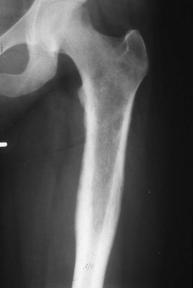
This is a plain radiograph that shows AP and lateral views of the distal lower extremity. The epiphyses are open, consistent with the patient’s age. There is a diffuse process involving the tibia from the distal epiphysis to the proximal metaphysis. The appearance suggests an aggressive process due to the “moth-eaten” appearance. The periosteum appears to be significantly lifted from the bone – this can be seen, particularly in young children, resulting in subperiosteal abscesses.
|
Sections show fragments of bone with osteonecrosis – the lacunae do not have osteocytes. There is an associated brisk acute inflammatory infiltrate composed primarily of neutrophils. Plasma cells are present in the fibrotic areas. Dark purple granular bacterial colonies are focally present.
|
Radiology image questions:
- Describe the plain radiograph. Take a screenshot and annotate: the tibia, fibula, growth plate and the area of the lesion.
- Does this appear to be an indolent or aggressive lesion?
VM image questions:
- Describe the microscopic findings.
- Take a screenshot and annotate: cortical bone, osteonecrosis, woven bone, lamellar bone and bacterial colonies.
- Which types of inflammatory cells are present? Support your answer with an annotated screenshot.
510-2. Which of the following is the most likely etiology of this condition in this patient?
- Anti-citrullinated peptide antibodies
- Corticosteroid use
- Escherichia coli infection
- Morbid obesity
- Staphylococcus aureus
510-3. Which of the following is a rare complication of this condition?
- Chronic renal failure
- Osteoporosis
- Osteosarcoma
- Paget disease
- Squamous cell carcinoma
510-4. A 15-year-old boy is diagnosed with osteomyelitis of the left tibia. Cultures are positive for Salmonella typhi. This patient most likely has which of the following?
- Compound fracture
- Hyperparathyroidism
- Osteomalacia
- Sickle cell disease
- Vitamin D deficiency
CASE NUMBER 223
Clinical History: A 13-year-old boy presented to his pediatrician with a 6-week history of left knee pain and swelling. A plain radiograph was obtained.
223-1a. Based on the clinical history and findings thus far, what is the differential diagnosis?
Image Gallery:
This plain radiograph shows open epiphyses, consistent with the patient’s age. There is a destructive metaphyseal tumor with a moth-eaten appearance (ie. aggressive features). A soft tissue density that appears to be partially calcified can be appreciated.
|
Webslide 0301, developing finger, long. sect.
[ ImageScope]
[ WebScope]
The bones shown in Webslide 301 (distal end of a metacarpal, and the 1st, 2nd, and part of the 3rd phalangeals) are still undergoing the process of endochondral ossification, but there are many general features of long bones that can be appreciated. The shaft of each bone (the diaphysis) consists of a rigid cylinder of compact bone ( example) surrounding a hollow marrow cavity ( example). Within the marrow cavity are hematopoietic cells and some adipocytes amongst spicules of trabeculated, or spongy, bone. As most long bones mature, the hematopoietic cells are replaced by the adipocytes and the marrow transitions from being 'red' (hematopoietic) to 'yellow' (fatty).
Outside of the cylinder of compact bone is a connective tissue sheath known as the periosteum. The periosteum is further subdivided into an inner, cellular periosteum (close to the bone), which is less dense in nature and made up of fibroblasts and osteoprogenitor cells and an outer, fibrous periosteum made up of dense, irregular connective tissue. The periosteum adheres to the bone by way of Sharpey's fibers that extend from the fibrous layer into the compact bone (example).
The articular surfaces at the ends of the bones (the epiphyses) are comprised of hyaline cartilage. Active growth plates (or epiphyseal plates) may be observed at the proximal ends of the phalanges (example) and the distal end of the metacarpal bone (example) whereas the cartilage at the distal ends of the phalanges exhibits much less proliferation (example), which is consistent with the general pattern of growth observed these and other long bones.
050_HISTO_40X, fibula, cross sect.
[ImageScope]
[WebScope]
This is a cross section of a fibula from an adolescent primate and therefore exhibits characteristics of more mature bone. The marrow cavity still contains a great deal of hematopoietic cells and would therefore be considered ‘red’ --however, note that there is a significant amount of adipose tissue that will increase over time. The shaft of the fibula consists of compact bone which is organized into inner (example) and outer (example) circumferential lamella, made up of bone laid down in concentric layers. Sandwiched between the inner and outer lamellae are collections of osteons, or Haversian systems (example). Within the bone, you can see the individual osteocytes within their lacunae and the small canaliculi through which the osteocytes extend processes to communicate with each other. The periosteum is relatively thin and consists primarily of an outer fibrous layer of dense irregular connective tissue. Outside the periosteum are fibers of skeletal muscle associated with the extensor and flexor compartments of the leg.
|
Clinical history, part 2 (click here to open)
CASE NUMBER 161
[DigitalScope]
Clinical History: A 16-year-old boy presented to the pediatrician with a 1-month history of pain and swelling of the left hip. He told the pediatrician that he had been struck there by a baseball four months earlier. Plain radiography was performed, followed by a biopsy and limb-salvage surgery.
Neoadjuvant chemotherapy was given prior to limb-salvage surgery.
Image Gallery:
Review Bone Histology
Webslide 0301, developing finger, long. sect.
[ DigitalScope]
The bones shown in Webslide 301 (distal end of a metacarpal, and the 1st, 2nd, and part of the 3rd phalangeals) are still undergoing the process of endochondral ossification, but there are many general features of long bones that can be appreciated. The shaft of each bone (the diaphysis) consists of a rigid cylinder of 'compact' or 'cortical' bone surrounding a hollow marrow cavity. Within the marrow cavity are hematopoietic cells and some adipocytes amongst spicules of 'trabeculated,' or 'spongy,' bone. As most long bones mature, the hematopoietic cells are replaced by the adipocytes and the marrow transitions from being 'red' (hematopoietic) to 'yellow' (fatty).
in addition to the terms 'compact' and 'spongy,' bone can also be classifed as 'lamellar' (mature) or 'woven' (immature). As the name implies, woven bone consits of many osteocytes within an irregularly organized ossified matrix that has a woven appearance. As the bone matures, it becomes organized into lamellar bone that features lamella of sparse osteocytes separated by distinct layers of ossified matrix.
Outside of the cylinder of compact bone is a connective tissue sheath known as the periosteum. The periosteum is further subdivided into an inner, cellular periosteum (close to the bone), which is less dense in nature and made up of fibroblasts and osteoprogenitor cells and an outer, fibrous periosteum made up of dense, irregular connective tissue. The periosteum adheres to the bone by way of Sharpey's fibers that extend from the fibrous layer into the compact bone.
The articular surfaces at the ends of the bones (the epiphyses) are comprised of hyaline cartilage. Active growth plates (or epiphyseal plates) may be observed at the proximal ends of the phalanges and the distal end of the metacarpal bone whereas the articular cartilage at the distal ends of the phalanges exhibits much less proliferation, which is consistent with the general pattern of growth observed these and other long bones.
050_HISTO_40X, fibula, cross sect.
[DigitalScope]
This is a cross section of a fibula from an adolescent primate and therefore exhibits characteristics of more mature bone. The marrow cavity still contains a great deal of hematopoietic cells and would therefore be considered ‘red’ --however, note that there is a significant amount of adipose tissue that will increase over time. The shaft of the fibula consists of compact bone which is organized into inner and outer circumferential lamella, made up of bone laid down in concentric layers. Sandwiched between the inner and outer lamellae are collections of osteons, or Haversian systems. Within the bone, you can see the individual osteocytes within their lacunae and the small canaliculi through which the osteocytes extend processes to communicate with each other. The periosteum is relatively thin and consists primarily of an outer fibrous layer of dense irregular connective tissue. Outside the periosteum are fibers of skeletal muscle associated with the extensor and flexor compartments of the leg.
|
The radiograph shows a lytic, infiltrative lesion in the proximal half of the femur. There are reactive periosteal changes in the proximal diaphysis with an onion-skin appearance. The MRI shows the tumor extending into the soft tissue. Cross section through the resected femoral head shows a soft tan white tumor with focal hemorrhage and necrosis. There is extensive soft tissue invasion.
|
A striking feature is the monotony of the cells with round to oval vesicular nuclei and poorly defined, scanty eosinophilic cytoplasm. There are a moderate number of mitoses, but tumor giant cells and pleomorphism are conspicuously absent. The tumor cells are arranged around scanty, very vascular, fibrous stroma in a trabecular fashion, sometimes resembling rosette formation. In between these cords the tumor cells seem to line empty spaces. Histologically it is quite difficult to distinguish this tumor from a neuroblastoma.
|
161-1. What is the differential diagnosis?
161-2. Which of the following is most accurate concerning this entity?
- Cellular pleomorphism is usually marked
- Most patients are older than 20 years at diagnosis
- Plain radiography often shows an epiphyseal lytic lesion
- Skeletal muscle differentiation is common
- There is a recurrent chromosomal translocation in most cases
161-3. Which of the following is considered part of the spectrum of this tumor?
- Fibrous dysplasia
- Giant cell tumor of bone
- Neuroblastoma
- Primitive neuroectodermal tumor
- Small cell osteosarcoma
CASE NUMBER 227
[DigitalScope]
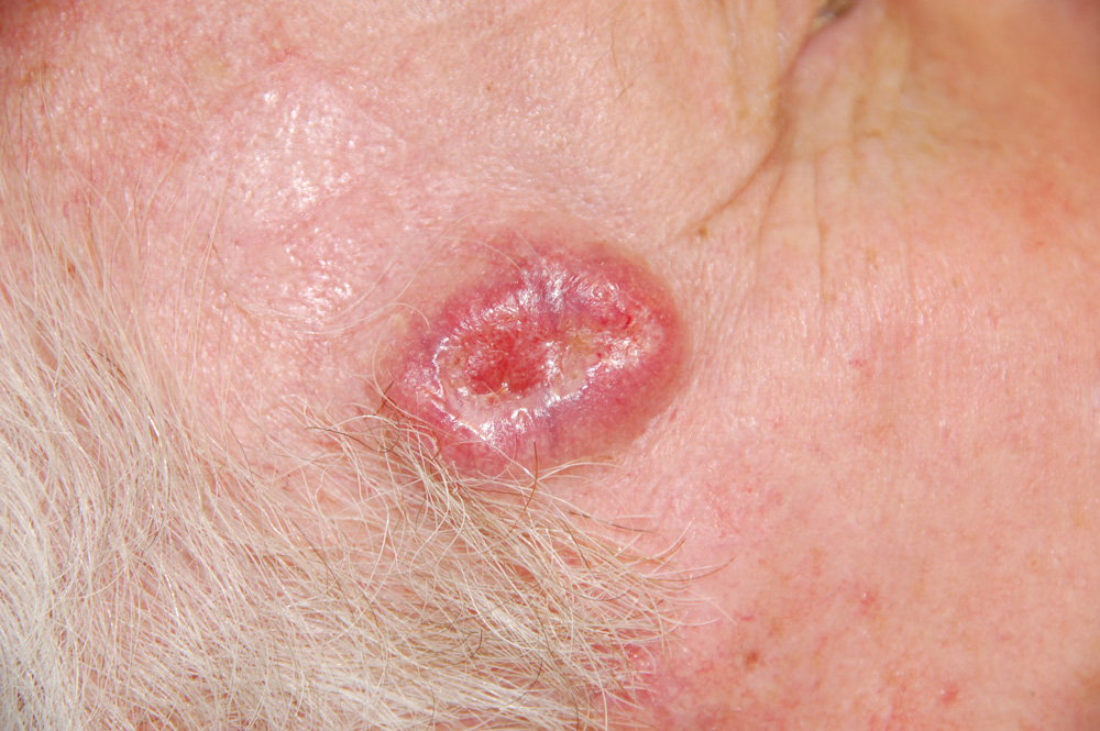
Clinical History: A 62-year-old man presented to his primary care physician with a 3-year history of a slowly growing lesion on his right temple. The lesion was excised. Gross and microscopic images are provided.
227-1. What is the differential diagnosis?
Image Gallery:
Review Skin Histology
Norm No. 15 Skin
[DigitalScope]
Skin consists of keratinizing stratified squamous epithelium. The keratin layer is eosinophilic. Nuclei are not present. Beneath the keratin layer is a layer of flat epithelial cells with small pyknotic nuclei. Cells are regular and not crowded. These keratinocytes rest upon a layer of basal epithelium and a thin basement membrane. Beneath the epidermis is the dermis which consists of loose connective tissue. Within the dermis are sweat and sebaceous glands. Some slides contain hair follicles. There is no inflammation. The blood vessels are patent and do not contain thromboemboli.
|
There was a well circumscribed, firm, elevated mass 1cm in diameter. The overlying skin was intact but smooth and has a pearly, pink appearance. There is central umbilication.
|
There are nests of darkly staining basophilic cells in the upper dermis with peripheral palisading. There is little cytoplasm, and the cytoplasmic borders are poorly defined. Nuclei are small and are round or oval, and occasional apoptotic bodies are noted. These tumors may arise from the basal cell layer of the epidermis or from dermal appendages. In this particular tumor there is an occasional round mass of keratin which is not usually seen in basal cell carcinomas. Note the loose, myxoid stroma about the tumor cells with some artefactual clefts.
|
227-2. Which of the following is a characteristic histologic finding in this tumor?
- Eosinophilic, inclusion-like nucleoli and cytosolic melanin pigment
- Lichenoid inflammatory infiltrate
- Loose myxoid stroma around the tumor cells with artefactual clefts
- Nests of small cells containing neurosecretory granules and cytokeratin 20
- Suprabasal acantholytic blister
227-3. Which of the following is true regarding the natural history of this tumor?
- Frequently metastasizes to lung
- Initially indolent, then widespread metastasis
- Locally destructive, but rarely metastasizes
- Metastasizes to regional lymph nodes
- Typically spreads along nerves and invades blood vessels
227-4. Which of the following is most strongly correlated with development of this tumor?
- BRAF mutation
- Cigarette smoking
- Drug exposure
- Sun exposure
- Varicella zoster infection
MUSCULOSKELETAL Review Items
Key Vocabulary Terms (click here to search any additional terms on Stedman's Online Medical Dictionary)
LEARNING OBJECTIVES
Goal 1: Nonneoplastic Disorders of the Musculoskeletal System
Apply knowledge of histology, immunology, microbiology, and biological and molecular alterations to discuss clinical presentation, biological behavior, morphological appearance, and natural history of nonneoplastic disorders of bones, joints, and skeletal muscle.
Objective 1: Osteomalacia and Rickets
Compare and contrast osteomalacia and rickets with respect to pathogenesis and clinicopathologic features.
Objective 2: Osteomyelitis
Discuss the pathogenesis of osteomyelitis, including predisposing factors, organisms involved, morphologic appearance, and complications.
Objective 3: Osteoporosis
Distinguish primary from secondary osteoporosis in terms of etiology, pathogenesis, and morphology.
Objective 4: Pathologic Fracture
Compare and contrast pathologic versus nonpathologic fractures including the potential for healing.
Objective 5: Paget Disease
Discuss the clincopathologic changes of Paget Disease including the histologic phases, genetic changes, and complications of this disorder.
Objective 6: Gout and Pseudogout
Compare and contrast gout and pseudogout in terms of epidemiology, clinical presentation, laboratory values, pathologic findings and treatment.
Objective 7: Arthritis
Compare and contrast rheumatoid and osteoarthritis including the etiology, pathogenesis, and morphology of each.
Goal 2: Bone Neoplasia
Apply knowledge of the molecular basis of neoplasia to describe the clinical presentation, biologic behavior, morphologic appearance, classification, diagnosis, prognosis, and targeted therapy of bone neoplasms.
Objective 1: Categories of Bone Tumors
Describe examples of bone forming, cartilage forming, and other common bone tumors including the clinicopathologic features, radiological features, treatment, and prognosis of each.
Objective 2: Bone-Forming Sarcomas
Describe the most common benign and malignant bone forming tumors in terms of epidemiology, clinical presentation, radiologic findings, histologic features, treatment, and prognosis.
Objective 3: Cartilage-Forming Sarcomas
Describe the most common benign and malignant cartilaginous tumor of bone in terms of epidemiology, clinical presentation, radiologic findings, histologic features, treatment, and prognosis.
Objective 4: Metastatic Tumors
Describe the tumors that commonly metastasize to bone, the radiologic manifestations of metastatic lesion involving bone, and the difference between osteoblastic and osteolytic metastases.
Objective 5: Soft-Tissue Tumors
Describe the common benign and malignant soft tissue tumors including the genetic contribution to tumor development and progression.
MDERMATOLOGIC Review Items
Key Vocabulary Terms (click here to search any additional terms on Stedman's Online Medical Dictionary)
LEARNING OBJECTIVES
Skin
Goal 1: Classification of Skin Disease
Apply knowledge of histology, cell biology, inflammation, and neoplasia to an understanding of the clinical presentation, biologic behavior, morphologic appearance, and classification of diseases of the skin.
Objective 1.1: Pathophysiology of Changes in the Skin
Describe the pathophysiologic basis for changes in the color, surface texture and swelling of skin.
Objective 1.2 Nomenclature of Skin Lesions
Compare and contrast the different types of macroscopic and microscopic lesions.
Goal 2: Infections of the Skin
Apply knowledge of the anatomic and immunologic structure of the skin to discuss the role of skin in protecting against direct invasion of skin and appendages by pathogens.
Objective 2.1: Cutaneous Infections
Describe common bacterial, viral, fungal, and parasitic agents that may cause cutaneous infections and the particular sites that they infect, and morphologic features and complications of these infections.
Objective 2.2 Acute Inflammatory Dermatoses
Compare and contrast the major acute inflammatory dermatoses including urticaria, acute eczematous dermatitis and erythema multiforme in terms of risk factors, etiology, clinical presentation, pathologic features, natural history, prognosis and treatment.
Objective 2.3 Chronic Inflammatory Dermatoses
Compare and contrast the major chronic inflammatory dermatoses including psoriasis, lichen planus and lichen simplex chronicus in terms of risk factors, etiology, clinical presentation, pathologic features, natural history, prognosis and treatment.
Goal 3: Immune-Related Disorders of the Skin
Apply knowledge of basic concepts in immunopathology and the key immunologic functions of components of the skin to understand the pathologic basis of disease caused by reactivity to exogenous agents versus immunologically driven disease with a genetic component.
Objective 3.1: Immune Diseases of the Skin
Describe the clinical features, pathophysiology and pathologic findings for the following immunologically driven diseases with a genetic component: pemphigus (vulgaris and foliaceus), bullous pemphigoid, dermatitis herpetiformis
Goal 5: Skin Neoplasia
Apply knowledge of the molecular basis of neoplasia to an understanding of the clinical presentation, biologic behavior, morphologic appearance, classification, diagnosis, prognosis, and therapy of benign and malignant skin neoplasms.
Objective 5.1: Benign Skin Neoplasms
Compare and contrast seborrheic keratosis and actinic keratosis in terms of clinical presentation, etiology, pathologic findings and risk of malignant transformation.
Objective 5.2: Malignant Skin Neoplasms
Describe the clinical presentation, precursor lesions, risk factors, genetic basis, hereditary cancer syndromes and pathologic features of the following skin cancers: basal cell carcinomas, squamous cell carcinoma, and melanoma.
Objective 5.3: Sun Exposure
Explain the role of ultraviolet light and other environmental factors in development of various skin cancers.
|