CASE NUMBER 25
[ImageScope] [WebScope]
Clinical History: A 9-year-old girl presented to her pediatrician with a 6-week history of painful, swollen joints and recent onset dyspnea. Physical exam revealed a temperature of 38C, cervical lymphadenopathy and pansystolic and diastolic murmurs. Laboratory analysis was positive for serum antibodies to streptolysin O. Although she was admitted to the hospital, she died of progressive heart failure.
Image Gallery:
(Summary of Gross Findings - click here)
Her heart weighed 380 grams (normal for this age is about 115 grams). The pericardium was covered with a fibrinous exudate. The left ventricle was dilated and the myocardium was flabby. The mitral valve was slightly thickened as were the chordae tendinae. There was a MacCallum's patch in the left atrium. The liver weighed 780 grams (normal 750 grams). There was centrilobular congestion ("nutmeg liver").
|
(Summary of Microscopic Findings - click here)
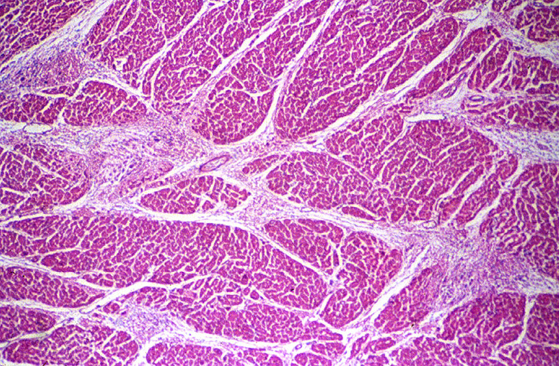
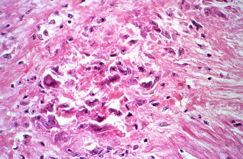
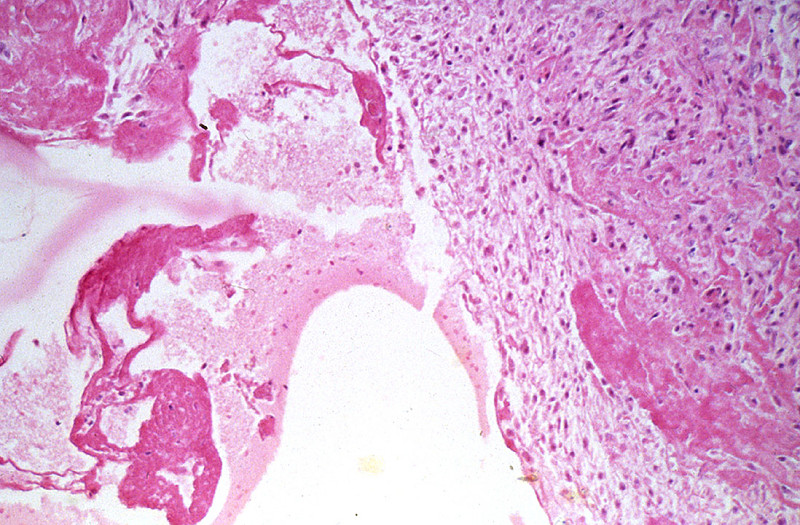
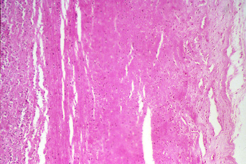
The section includes the entire thickness of the myocardium and is taken through the mitral valve so that both left atrial and left ventricular myocardium are present in the section. The epicardium shows a prominent layer of fibrin on the surface; deep to this is young connective tissue with many capillaries, fibroblasts, and chronic inflammatory cells, i.e. granulation tissue. Thus, this is an organizing fibrinous pericarditis. There is also myocarditis present. The myocardial inflammation includes Aschoff bodies of different ages. Identify very early, intermediate and healed foci. The presence of Aschoff bodies indicates that this is a rheumatic myocarditis. Note also marked endocardial thickening due to inflammation and scarring, especially in the left atrium (MacCallum's patch).
|
(Review Normal Histology - click here)
Norm No. 13 Heart
[ImageScope] [WebScope]
Normal heart tissue sections demonstrate no evidence of fibrosis or hemorrhage. Cardiac myocytes have moderately sized centrally located nuclei. Normal myocytes are not brightly eosinphilic. Normally no inflammation is seen. Normal cardiac myocytes do not show hypertrophy.
|
25-1. Which of the following is the most likely diagnosis?
- Amyloidosis
- Carcinoid heart disease
- Nonbacterial thrombotic endocarditis
- Rheumatic heart disease
- Systemic lupus erythematosus
ANSWER
25-2. The microscopic images show macrophages with abundant cytoplasm and central round to oval nuclei with a central ribbon of chromatin that has been liked to “owl eyes” or “caterpillars”. Which of the following is the term for the pathognomonic cells in this disease?
- Anitschkow cells
- Aschoff bodies
- Lacunar cells
- Langerhans cells
- Reed-Sternberg cells
ANSWER
25-3. Which of the following is the proposed pathogenesis of this disease?
- Antibodies to streptococcus cross-react with heart antigens
- Bioactive products released by the tumor cause fibrosis
- Direct toxicity of catecholamines on cardiac myocytes
- Endocardial trauma acts as a nidus for sterile vegetations
- Mutations in sarcomeric proteins
ANSWER
25-4. Which of the following is the most common cause of mitral stenosis?
- Amyloidosis
- Carcinoid heart disease
- Nonbacterial thrombotic endocarditis
- Rheumatic heart disease
- Systemic lupus erythematosus
ANSWER
CASE NUMBER 195
[ImageScope] [WebScope]
Clinical History: A 65-year-old man who had been diagnosed with rheumatic heart disease when he was 35-years old presented to his primary care physician with a 3-month history of fever and chills and dyspnea. Physical exam revealed bilateral lower extremity pitting edema, splinter hemorrhages and a purpuric skin rash. He was admitted to the hospital but died.
Image Gallery:
(Summary of Gross & Lab Findings - click here)
Splinter hemorrhages were observed at digit tips. Blood cultures grew alpha-hemolytic streptococci. The alpha hemolysis on blood agar typical of Streptococcus viridans is shown. Gram stain shows gram positive cocci in chains.The heart weighed 400 grams. There was thickening of the mitral valve leaflets and the chordae tendineae. Many friable calcified pink-gray granular verrucae were present on the valve. In addition, a large vegetation of the same type was found on the left auricular endocardium.
|
(Summary of Microscopic Findings - click here)
The section represents a portion of mitral valve, left atrium and left ventricle. The valve is greatly thickened and damaged. It is infiltrated with acute and chronic inflammatory cells, and shows a zone of necrosis and fibrosis in the central portion of the valve. The myocardium shows slight focal fibrosis, and focal acute inflammatory infiltration in some sections.
|
(Review Normal Histology - click here)
Norm No. 13 Heart
[ImageScope] [WebScope]
Normal heart tissue sections demonstrate no evidence of fibrosis or hemorrhage. Cardiac myocytes have moderately sized centrally located nuclei. Normal myocytes are not brightly eosinphilic. Normally no inflammation is seen. Normal cardiac myocytes do not show hypertrophy.
|
195-1. Which of the following is the most likely diagnosis?
- Bacterial endocarditis
- Carcinoid heart disease
- Libman-Sacks endocarditis
- Nonbacterial thrombotic endocarditis
- Viral endocarditis
ANSWER
195-2. In this patient, which of the following is the most likely causative organism?
- Group A streptococcus
- Neisseria meningitidis
- Staphylococcus aureus
- Staphylococcus epidermidis
- Viridans streptococcus
ANSWER
195-3. Involvement of which of the following valves most suggestive of IV drug abuse?
- All valves are equally affected
- Aortic valve
- Mitral valve
- Pulmonic valve
- Tricuspid valve
ANSWER
CASE NUMBER 31
[ImageScope] [WebScope]
Clinical History: A 45-year-old man presented to the emergency department with a 2-hour history of chest pain that radiated to both arms and his neck. Physical exam showed diaphoresis and blood pressure of 160/110 mmHg. He was treated with the diuretic furosemide; however, two days after the onset of his chest pain, he experienced a cardiac arrest and died.
Image Gallery:
(Summary of Gross Findings - click here)
The heart was slightly enlarged weighing 460g. There was severe atherosclerosis of all the major coronary arteries with a recent thrombotic occlusion of the proximal left anterior descending coronary artery. A recent transmural infarct was present in the left ventricle that involved the interventricular septum and the papillary muscle.
|
(Summary of Microscopic Findings - click here)
The slide includes a transmural section of the left ventricle. Nearly the entire section is involved by infarct. However, there is a thin rim (5 to 10 cell layers) of endocardial myocytes which have survived because of diffusion of oxygen and nutrients from the ventricular cavity. Other viable myocytes can be found around larger blood vessels within the section. The intense hypereosinophilia of the necrotic myocytes can best be appreciated by comparing the thin rim of lighter staining subendocardial myocytes with the deeper cells. Note also the karyolysis that is characteristic of coagulation necrosis. In some areas there is little inflammatory response. This observation is explained by microvascular necrosis which does not allow access of circulating leukocytes to these areas. In other areas, especially in the epicardial half of the infarct, there is an intense acute inflammatory response. Many intact neutrophils can be seen. In addition, there are many nuclear fragments from lysed neutrophils. Macrophage activity is not evident. These features of the inflammatory response indicate that the infarct was approximately three to four days old. Note also that the inflammation extends to the epicardial surface and that there are deposits of fibrin on the epicardium. This is called fibrinous pericarditis. The granular grey material seen within some blood vessels is barium sulfate, which was injected to permit post-mortem study of the coronaries by radiography.
|
(Review Normal Histology - click here)
Norm No. 13 Heart
[ImageScope] [WebScope]
Normal heart tissue sections demonstrate no evidence of fibrosis or hemorrhage. Cardiac myocytes have moderately sized centrally located nuclei. Normal myocytes are not brightly eosinphilic. Normally no inflammation is seen. Normal cardiac myocytes do not show hypertrophy.
|
31-1. Which of the following is the most likely diagnosis?
- Acute myocardial infarct
- Amyloidosis
- Bacterial myocarditis
- Dilated cardiomyopathy
- Hemorrhagic pericarditis
ANSWER
31-2. Which of the following was most likely the precipitating event in this patient’s disease?
- Brain stem herniation
- Fixed coronary obstruction
- Hypovolemic shock
- Occlusive thrombus
- Plaque disruption
ANSWER
31-3. Coronary artery bypass is a surgical intervention that can prolong life in patients with diseased arteries. Which of the following would be the best choice for bypass of an isolated coronary artery stenosis?
- Expanded polytetrafluoroethylene
- Left internal mammary artery
- Left saphenous vein
- Right internal mammary artery
- Right saphenous vein
ANSWER
31-4. Which of the following arteries was most likely occluded in this patient?
- Left anterior descending coronary artery
- Left circumflex coronary artery
- Right coronary artery
ANSWER
31-5. Which of the following sites is most vulnerable to ischemic injury in hypovolemic shock?
- Mid-myocardial, left ventricle
- Mid-myocardial, right ventricle
- Subendocardial, left ventricle
- Subendocardial, right ventricle
- Subepicardial, left ventricle
- Subepicardial, right ventricle
ANSWER
31-6. Which of the following microscopic features is most indicative of reperfusion of irreversibly damaged ischemic myocardium?
- Endothelial swelling
- Hypereosinophilic cytoplasm
- Necrosis with contraction bands
- Neutrophilic infiltrate
- Wavy fibers
ANSWER
CASE NUMBER 286
[ImageScope] [WebScope]
Clinical History: A 64-year-old man presented to the emergency department with a two-week history of worsening dyspnea and lower leg edema. He reported that he had two episodes of myocardial infarction three years earlier and that he had been subsequently diagnosed with congestive heart failure 8 months ago. While under observation, he experienced a fatal arrhythmia.
Image Gallery:
(Summary of Gross Findings - click here)
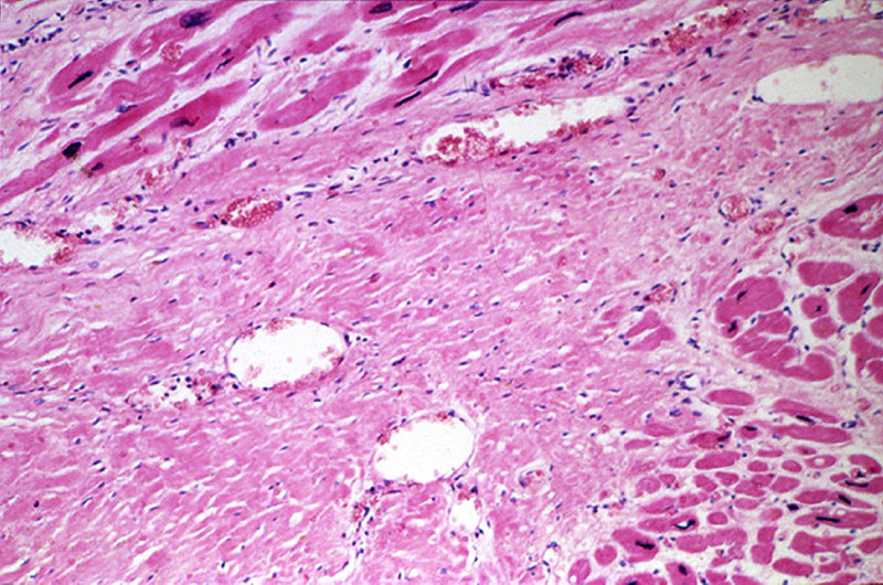
The heart was markedly hypertrophied (740 grams) and dilated. There was marked coronary atherosclerosis with an old occlusion of the left anterior descending vessel. A healed infarct involved the anteroseptal and apical region of the left ventricle. A mural thrombus which is not shown here covered much of the infarct.
|
(Summary of Microscopic Findings - click here)
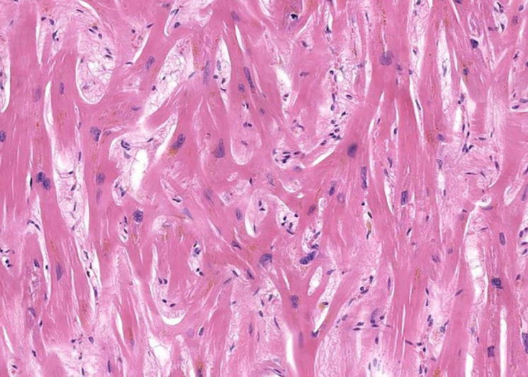
The slide includes a transmural section of the anterior free wall and anterior portion of the interventricular septum. There is a broad band of dense, highly collagenized scar tissue replacing the middle layer of myocardium; patchy scarring interspersed with hypertrophied cardiac myocytes is present on either side of this dense scar. The endocardium is markedly thickened and there is organizing mural thrombus between cardiac trabeculae and extending into the lumen. Some myocytes in the subendocardial layer show sarcoplasmic vacuolization, a chronic degenerative change termed "myocytolysis".
|
(Review Normal Histology - click here)
Norm No. 13 Heart
[ImageScope] [WebScope]
Normal heart tissue sections demonstrate no evidence of fibrosis or hemorrhage. Cardiac myocytes have moderately sized centrally located nuclei. Normal myocytes are not brightly eosinphilic. Normally no inflammation is seen. Normal cardiac myocytes do not show hypertrophy.
|
286-1. Which of the following is the most likely diagnosis?
- Acute myocardial infarct
- Amyloidosis
- Arrhythmogenic right ventricular cardiomyopathy
- Cor pulmonale
- Healed myocardial infarct
ANSWER
286-2. Based on these images, which of the following is the youngest age that the pictured lesion could be?
- 3 hours
- 3 days
- 3 weeks
- 3 months
- 3 years
ANSWER
286-3. In the setting of a myocardial infarct, which of the following is the time when there is the greatest risk of free wall rupture?
- 3 hours
- 3 days
- 3 weeks
- 3 months
- 3 years
ANSWER
286-4. Patients with posterior transmural infarcts are at greatest risk for which of the following?
- Aneurysm
- Conduction blocks
- Free wall rupture
- Infarct expansion
- Mural thrombi
ANSWER
CASE NUMBER 512
(no virtual slides for this case)
Clinical History: A 19-year-old man is brought to the emergency department by ambulance following a syncopal episode that occurred while he was playing in a varsity basketball game 30 minutes earlier. Physical examination reveals a harsh systolic ejection murmur, ECG demonstrates prominent Q waves and echocardiography reveals left ventricular hypertrophy and asymmetric septal hypertrophy.
Image Gallery:
512-1. Which of the following is the most likely diagnosis?
- Acute pericarditis
- Amyloidosis
- Hypersensitivity myocarditis
- Hypertrophic cardiomyopathy
- Rheumatic heart disease
ANSWER
512-2. Analysis of a cardiac biopsy would most likely show which of the following?
- Abundant eosinophils
- Apple-green birefringent material
- Distinct nuclear and ill-defined cytoplasmic inclusions
- Missense mutation in the b-myosin heavy chain gene
- Non-caseating granulomas
ANSWER
512-3. Which of the following is true regarding this disease?
- Etiology includes genetic and non-genetic causes
- In women, it is most common in late pregnancy
- Mutations in cytoskeletal proteins are seen in about 50% of cases
- Patients receiving doxorubicin are at increased risk
- The ejection murmur is due to ventricular outflow obstruction
ANSWER
512-4. Which of the following gross findings is commonly seen at autopsy in these patients?
- “Banana-like” configuration of the left ventricular cavity
- Bilateral atrial dilation
- Commisural fusion
- Firm, plaque-like endocardial fibrous thickenings
- Mitral annular calcification
ANSWER
CARDIOVASCULAR PATHOLOGY Review Items
Key Vocabulary Terms (click here to search any additional terms on Stedman's Online Medical Dictionary)
LEARNING OBJECTIVES
Absolutely critical information you must know to practice medicine is in bold font.
Important information that will be needed for routine patient care is in regular font.
Information about less common diseases that you may encounter in clinical practice and that will probably appear on examinations is in italics
-
List the most common forms of heart disease in the United States
- Contrast and compare the clinical and pathologic features of the following:
- high-output heart failure
- left-sided heart failure
- right-sided heart failure
- cor pulmonale
- Discuss cardiogenic shock in terms of:
- etiologic factors
- pathogenesis
- morphology
- stages
- clinical manifestations
- Discuss congenital heart disease in terms of:
- genetic and environmental factors
- types which result in:
- left-to-right vs. right-to-left shunts
- cyanotic vs. acyanotic disease
- types which come to medical attention in:
- infancy
- childhood
- adulthood
- Compare and contrast clinical and pathologic features of congenital heart disease:
- atrial septal defect (ASD)
- ostium primum
- ostium secundum
- venticular septal defect (VSD)
- tetralogy of Fallot
- endocardial cushion defects
- hypoplastic left heart syndrome
- patent ductus arteriosus (PDA)
- transposition of the great vessels
- coarctation of the aorta
- preductal
- postductal
- anomalous pulmonary venous return
- Compare and contrast clinical and pathologic features of the following
- endocarditis
- myocarditis
- pericarditis
- pericardial effusion
- cardiac tamponade
- pancarditis
- Compare and contrast the clinical and pathologic features of the following
- acute rheumatic fever.
- chronic rheumatic heart disease
- Compare and contrast the clinical and pathologic features of valvular heart disease
- calcific aortic stenosis
- aortic insufficiency
- mitral stenosis/insufficiency
- mitral valve prolapse
- mitral annular calcification
- tricuspid insufficiency
- pulmonic insufficiency
- infectious endocarditis
- List long term complications associated with prosthetic heart valves
- Compare and contrast the clinical and pathologic features of the following:
- dilated (congestive) cardiomyopathy
- hypertrophic cardiomyopathy (idiopathic hypertrophic subaortic stenosis (IHSS)
- restrictive cardiomyopathy
- endomyocardial fibrosis
- eosinophilic (Loeffler) endomyocarditis
- endocardial fibroelatosis
- Discuss coronary artery disease, in terms of:
- epidemiology
- risk factors
- etiologic factors
- pathogenesis
- complications
- Develop an understanding of acute coronary syndrome, relationship to plaque rupture and thrombosis, develop an understanding of the role of interventional cardiology and bypass surgery and why these treatments are used.
- Discuss myocardial infarct, in terms of:
- etiologic factors
- risk factors
- pathogenesis
- morphology
- evolution of morphologic changes with time
- correlation of morphologic distribution of infarct with site of coronary artery disease
- clinical, laboratory, and electrocardiography findings that change with time after the event
- complications, including expected timing after the event
- prognosis, and common causes of death with increasing time after the event
- Discuss sudden cardiac death, in terms of:
- causes
- relationship to arrhythmias
- cardiac morphology
- Discuss the following cardiac tumors
- myxoma
- rhabdomyoma
- lipoma
- metastatic
|