Case assignments by lab group and class day:
| |
|
|
| Labs 1, 2, & 3 |
|
|
| |
|
|
| Labs 4 & 5 |
|
|
| |
|
|
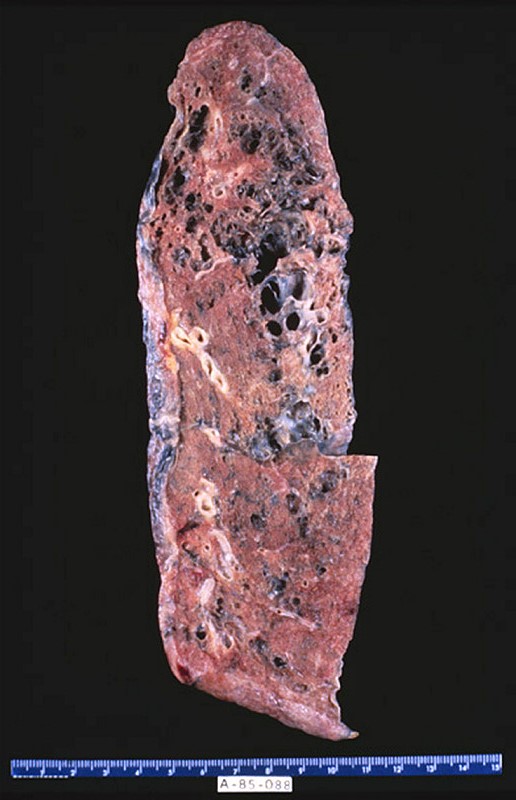
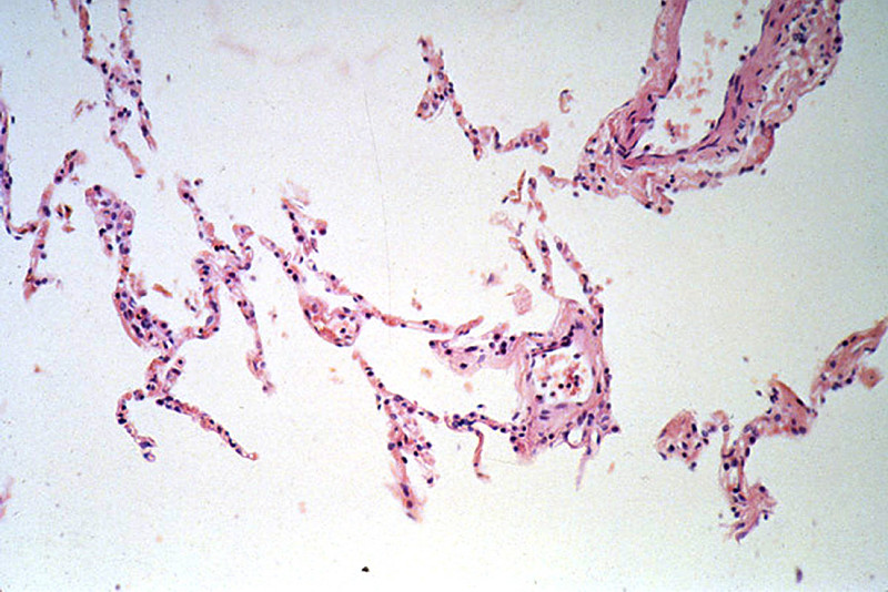
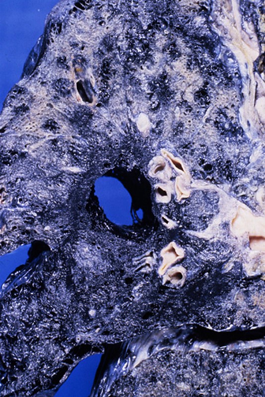
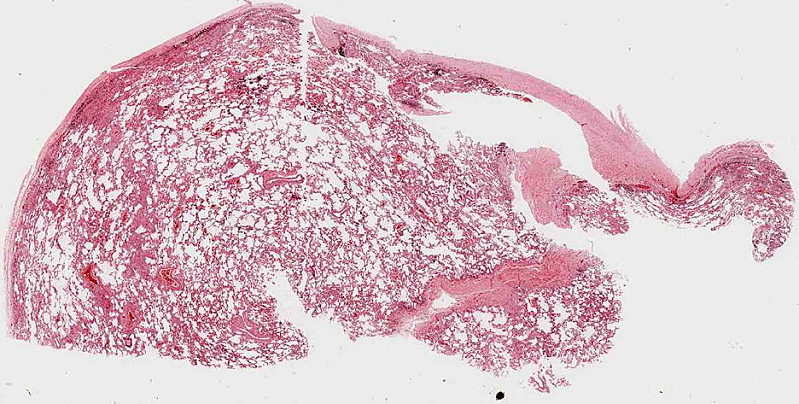
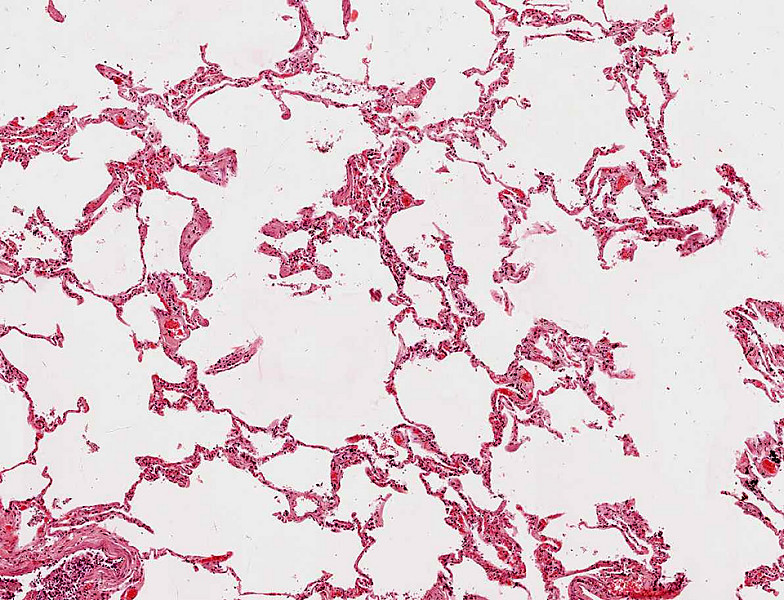
CASE NUMBER 92
[ImageScope] [WebScope]
Clinical History: A 65-year-old man presented to the emergency department with sudden onset dyspnea. He reported that he had been experiencing shortness of breath over the last ten years and that he had smoked 2 packs of cigarettes/day since he was 17. His medical history was significant for asthma as a child. He was admitted to the hospital for observation for worsening cardiac function, but experienced a myocardial infarction on the second day and died. Gross and microscopic images from the autopsy are provided.
Image Gallery:
(Summary of Gross Findings - click here)
The lungs were very large and upon palpation there was marked crepitation.
|
(Summary of Microscopic Findings - click here)
The alveolar spaces are distended and the alveolar septa are thickened by fibrous tissue. Numerous "free floating" alveolar septa are present, recognizable as pieces at alveolar wall not connected at either end to adjacent septa.
|
(Review Normal Histology - click here)
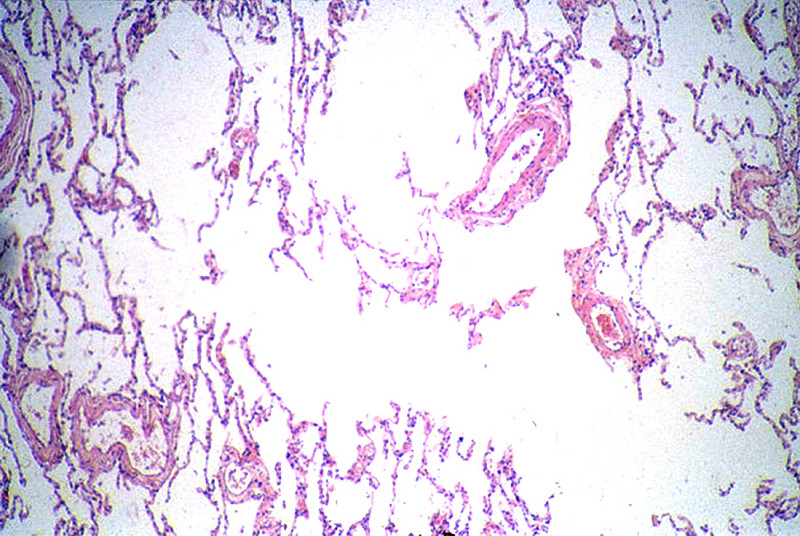
Norm No. 24 Lung
[ImageScope] [WebScope]
The primary function of the lung is gas exchange. Therefore, alveoli have thin walls lined by thin flat pneumocytes and endothelial cells. There is no thickening or fibrosis of the interstitium. The bronchioli are lined with basally oriented ciliated columnar epithelium. The bronchi are lined by similar epithelium. There are mucous glands within the submucosa. The bronchial smooth muscle is not hypertrophied. The pulmonary vessels are patent with no evidence of intimal thickening or muscular hyperplasia.
|
92-1. What is the differential diagnosis?
ANSWER
92-2. Which of the following is the most significant risk factor for this patient in the development of his disease?
- α1-antitrypsin deficiency
- Bronchial obstruction
- Cigarette smoking
- History of asthma
- Mutation in the PKD1 gene
ANSWER
92-3. Which of the following is considered the most likely mechanism by which lung tissue is damaged in this disease?
- Direct toxicity of tobacco smoke
- Free radical release generated by fibers
- IL-1 and TNF elaboration by macrophages
- Protease release by neutrophils
- Type III hypersensitivity
ANSWER
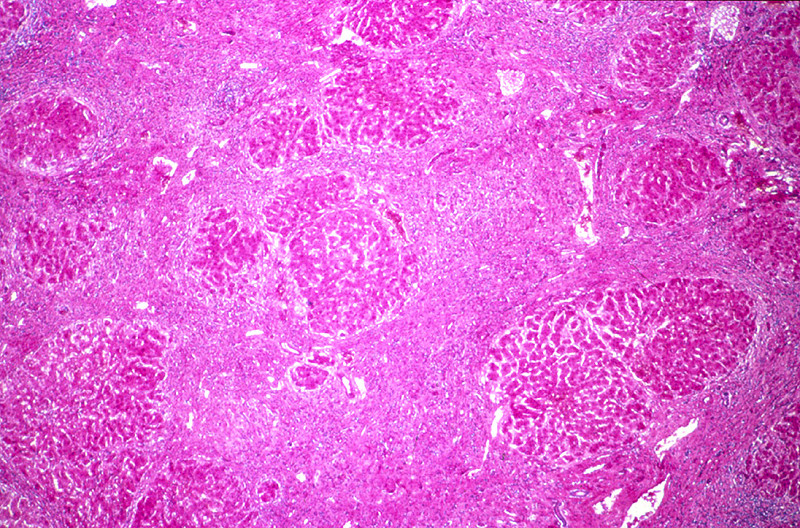
CASE NUMBER 81
[ImageScope] [WebScope]
Clinical History: A 62-year-old alcoholic man presented to his primary care physician with a 3-month history of increased abdominal girth. Physical exam revealed telangiectasias and fluid wave of the abdomen. Laboratory findings showed elevated aminotransferase and decreased serum albumin; no other significant findings were noted. He was admitted to the hospital for further evaluation, but began retching violently with extensive hematemesis. Volume resuscitation failed and the patient died.
Image Gallery:
(Summary of Gross Findings - click here)
The liver weighed 1800 grams. The entire organ was uniformly composed of nodules about 0.5 cm in diameter, each surrounded by fibrous tissue. The organ was jaundiced and firm.
|
(Summary of Microscopic Findings - click here)
The usual architecture present in the liver has been completely disrupted by the bands of connective tissue. In these bands one sees chronic inflammatory cells, mainly lymphocytes and other mononuclear cells. There is some proliferation of the bile ductules.
|
(Review Normal Histology - click here)
Norm No. 3 Liver
[ImageScope] [WebScope]
The liver is the organ that metabolizes nutrients received from the digestive tract. These nutrients and processed by tissue hepatocytes which are large polygonal cells. The hepatocyes are separated by portal triads. The triads consist of an artery, a vein and a bile duct. The bile duct is lined by cuboidal epithelium. The artery has a muscular wall and a flat endothelial lining. The sinuses are well defined and contain a small amount of blood.
|
81-1. What is the differential diagnosis?
ANSWER
81-2. Which of the following is the main enzyme involved in alcohol metabolism?
- Acetaldehyde dehydrogenase
- Alcohol dehydrogenase
- Catalase
- Cytochrome P450
- Glutathione peroxidase
ANSWER
81-3. Moderate intake of alcohol is most closely associated with which of the following?
- Bitot spots
- Hepatic adenoma
- Leptin secretion
- Nasal ulcers
- Steatosis
ANSWER
81-4. Which of the following is the mechanism that leads to the finding in question 3?
- Catalase levels are insufficient for cellular needs
- Decreased NAD+ levels impair fatty acid oxidation
- Endotoxin release stimulates TNF secretion
- Metabolism of ethanol produces reactive oxygen species
- Mucosal epithelium shifts to a squamous morphology
ANSWER
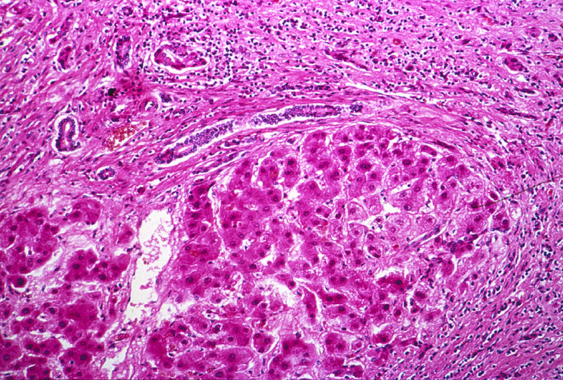
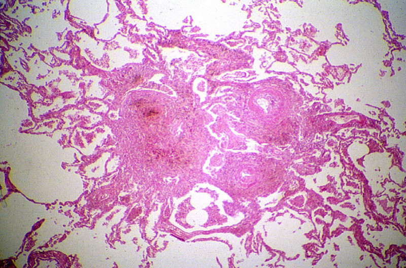
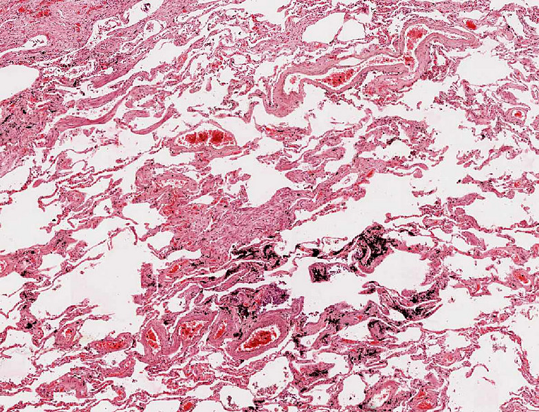
CASE NUMBER 98
[ImageScope] [WebScope]
Clinical History: A 70-year-old man presented to the emergency department with sudden worsening of his dyspnea. He reported that over the last 6 months he had been experiencing dyspnea, orthopnea, paroxysmal nocturnal dyspnea and ankle edema. Further questioning revealed that he worked in a rock quarry as a crusher for 18 years. He was admitted to the hospital for congestive heart failure, but died suddenly of a massive acute myocardial infarction.
Image Gallery:
(Summary of Gross and Lab Findings - click here)
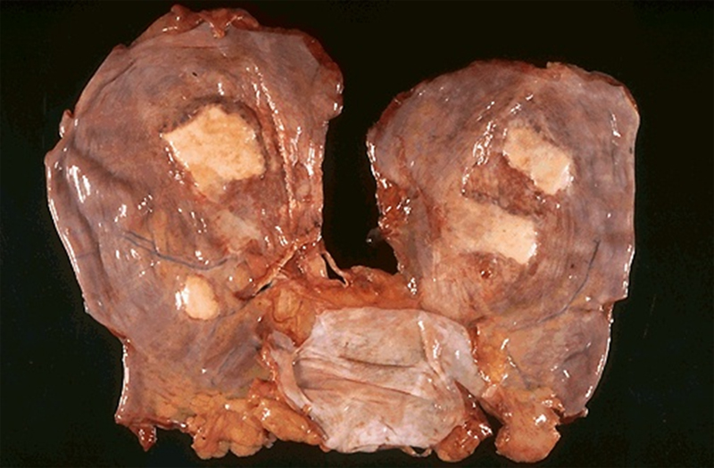
Both lungs were heavy, 900 grams for the left lung and 1120 grams for the right one. The lung was slate gray and firm.
|
(Summary of Microscopic Findings - click here)
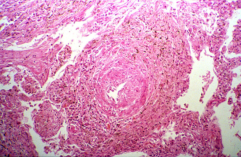
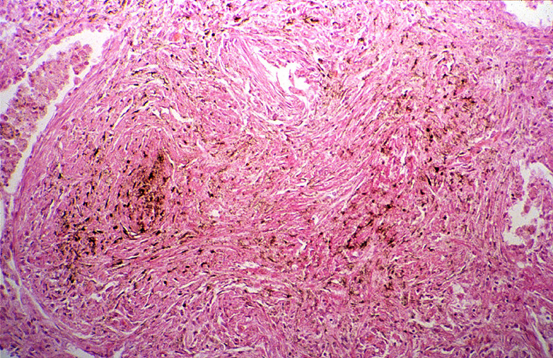
In the left upper part of the section the characteristic lesions of silicosis are seen. There are masses of fibrous tissue which form concentric lamination around the blood vessels, and stellate scars when the fibrosis extends to the adjacent alveolar septa. A large amount of coal-black pigment and small crystals are found in the macrophages and the scar tissue. These crystals are best seen under polarized light. In advanced lesion the fibrous nodules become confluent as seen in the lower right part of this section. Notice the presence of arteriosclerosis and organizing thrombi in the pulmonary arteries and chronic inflammation of bronchi.
|
(Review Normal Histology - click here)
Norm No. 24 Lung
[ImageScope] [WebScope]
The primary function of the lung is gas exchange. Therefore, alveoli have thin walls lined by thin flat pneumocytes and endothelial cells. There is no thickening or fibrosis of the interstitium. The bronchioli are lined with basally oriented ciliated columnar epithelium. The bronchi are lined by similar epithelium. There are mucous glands within the submucosa. The bronchial smooth muscle is not hypertrophied. The pulmonary vessels are patent with no evidence of intimal thickening or muscular hyperplasia.
|
98-1. What is the differential diagnosis?
ANSWER
98-2. Which of the following sites is most frequently affected by this disease?
- Bronchial mucosa
- Lower lobes of lungs
- Pleura
- Pulmonary arteries
- Upper lobes of lungs
ANSWER
98-3.Which of the following is the most common etiologic agent in this disease?
- Asbestos
- Carbon
- Cigarette smoke
- Micropolyspora faeni
- Quartz
ANSWER
98-4. Which of the following cells is most strongly implicated in the development of this disease?
- Endothelial cells
- Macrophages
- Plasma cells
- Smooth muscle cells
- T lymphocytes
ANSWER
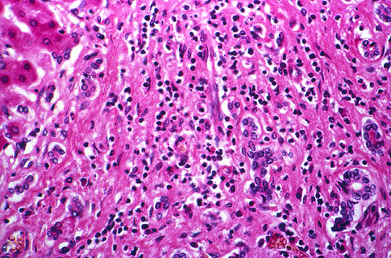
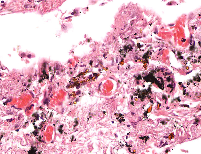
CASE NUMBER 560
[WebScope] [ImageScope]
Clinical History: A 72-year-old man was found dead in his home by his wife. She told the medical examiner that he was a retired shipyard worker who had smoked 1 pack of cigarettes/day since he was 19. Autopsy revealed that the cause of death was myocardial infarction.
Image Gallery (slide specimen and images courtesy of the University of Michigan):
(Summary of Gross Findings - click here)
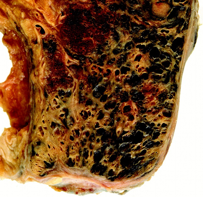
This is a section of the diaphragm showing the pleural side. There are dense white fibrocalcific plaques. On cut section, the surfaces of the lungs manifested diffuse fibrosis with extensive 'honeycombing.'
|
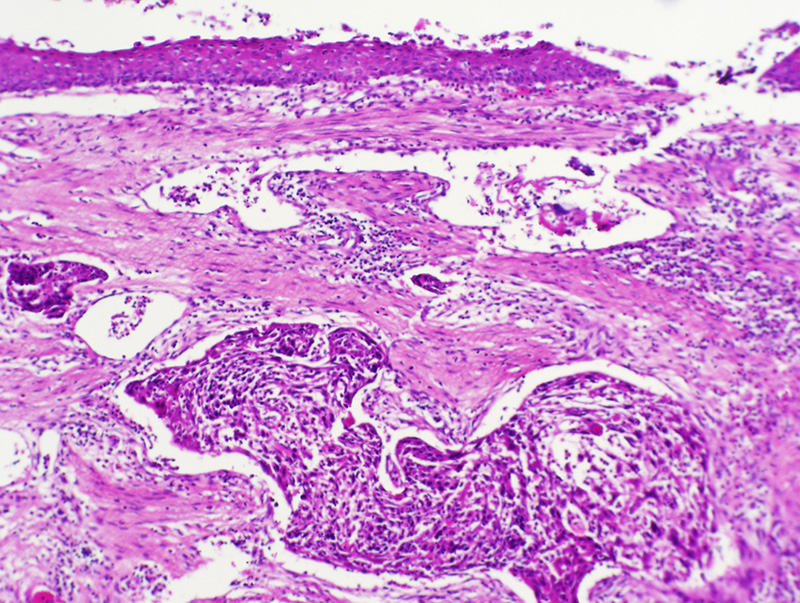
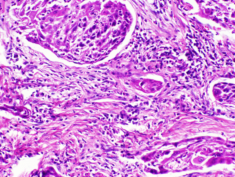
(Summary of Microscopic Findings - click here)
Many of the alveolar walls contain more nuclei than usual and there are detached fragments of alveolar septa. Small deposits of anthracotic pigment are also evident. Many of the alveolar septa are thickened by fibrosis. Within these areas of fibrosis are golden brown beaded or 'knobby' structures.
|
(Review Normal Histology - click here)
Norm No. 24 Lung
[ImageScope] [WebScope]
The primary function of the lung is gas exchange. Therefore, alveoli have thin walls lined by thin flat pneumocytes and endothelial cells. There is no thickening or fibrosis of the interstitium. The bronchioli are lined with basally oriented ciliated columnar epithelium. The bronchi are lined by similar epithelium. There are mucous glands within the submucosa. The bronchial smooth muscle is not hypertrophied. The pulmonary vessels are patent with no evidence of intimal thickening or muscular hyperplasia.
|
560-1. What is the differential diagnosis?
ANSWER
560-2. What are the golden-brown objects seen in the microscopic images?
- Beryllium fragments
- Charcot-Leyden crystals
- Ferruginous bodies
- Fungal spores
- Hyaline membranes
ANSWER
560-3. Patients with this disease are at greatest risk for developing which of the following complications?
- Acute interstitial pneumonia
- Bronchogenic carcinoma
- Diffuse alveolar damage
- Hypersensitivity pneumonitis
- Sarcoidosis
ANSWER
560-4. Which of the following pulmonary function test results is consistent with this patient’s diagnosis?
- Decreased forced vital capacity
- Decreased maximal expiratory flow rate
- Increased forced expiratory volume at 1 second
- Increased residual volume
- Increased total lung capacity
ANSWER
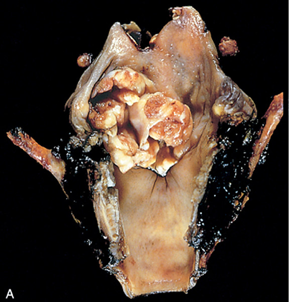
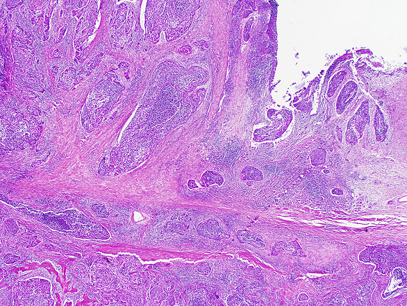
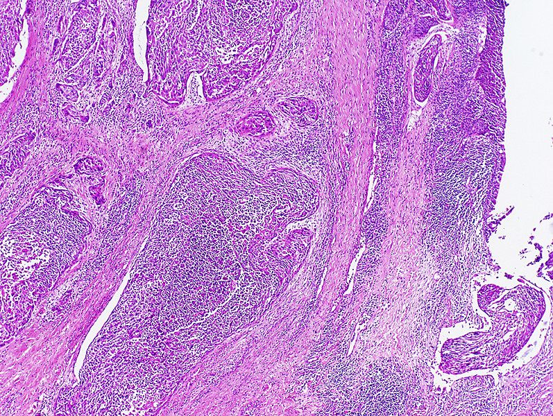
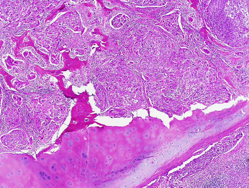
CASE NUMBER 154
[ImageScope] [WebScope] [DigitalScope]
Clinical History: A 64-year-old man presented to his primary care physician with a two-month history of severe dysphagia to solids and increasing dysphagia to liquids. He reported that he had lost approximately twenty pounds over the last six months. Additional findings elicited in his history and physical include 1) 50-pack year smoking history; 2) hepatitis B infection; and 3) work in a shipyard. Upper endoscopy revealed an ulcerated mass at the level of the vocal cords. A laryngectomy was performed.
Image Gallery:
(Summary of Gross Findings - click here)
An ulcerated tumor involved the left true and false vocal cords and extended across the midline.
|
(Summary of Microscopic Findings - click here)
The mucosa is partially columnar, representing the ventricular mucosa. There is a transition to squamous mucosa with marked nuclear pleomorphism. The abnormal squamous cells extend into the underlying stroma where the cells keratinize. Focal necrosis and an inflammatory infiltrate are present.
|
(Review Normal Histology - click here)
Slide 307 - Larynx
[ImageScope] [WebScope]
The larynx is a passageway for air between the oropharynx and trachea which also functions in the production of sound. It is lined by pseudostratified columnar epithelium (see left hand side of the slide), stratified squamous epithelium (covering the vocal cords near the middle of the slide), and stratified columnar epithelium between these epithelial types. Seromucous glands are present underneath the epithelium on both sides of the slide. Identify the vocal cord as a mucoal fold covered by stratified squamous epithelium and containing abundant skeletal muscle. Superior to the vocal fold (to the right of the focal fold on this slide) is the vestibular fold (aka "false" vocal fold), which is lined by respiratory epithelium. Notice the cartilage framework, within which there is some ossification as occurs as part of the aging process (this specimen is from a 62-year-old).
|
154-1. What is the differential diagnosis?
ANSWER
154-2. Which of the following risk factors is MOST closely linked to this disease?
- Asbestos exposure
- Copper deficiency
- Gastroesophageal reflux disease
- Hepatitis B infection
- Tobacco smoking
ANSWER
154-3. Recent studies have correlated HPV infection with oropharyngeal squamous cell carcinoma. Which of the following accounts for the carcinogenesis of high-risk HPV infection?
- Activation of cyclin-dependent kinase inhibitors
- Amplification of the MYC oncogene
- Displacement of E2F transcription factors from RB
- Inactivation of cyclin-dependent kinases
- Stabilization of p53
ANSWER
ENVIRONMENTAL PATHOLOGY Review Items
Key Vocabulary Terms (click here to search any additional terms on Stedman's Online Medical Dictionary)
LEARNING OBJECTIVES
Absolutely critical information you must know to practice medicine is in bold font.
Important information that will be needed for routine patient care is in regular font.
Information about less common diseases that you may encounter in clinical practice and that will probably appear on examinations is in italics
- Discuss the following in terms of role in indoor vs. outdoor air pollution:
- List the various substances found in cigarette smoke and their health effects.
- Discuss the effects and clinical significance of:
- active tobacco smoke
- passive (sidestream) tobacco smoke
- smokeless tobacco
- Outline the basic pathogenesis of pneumoconioses.
- Compare and contrast the following pneumoconiosis in terms of types of occupational exposure, pathogenesis and clinical course
- Compare coal workers' pneumoconiosis with simple asymptomatic anthracosis.
- Discuss Caplan syndrome in relation to coal workers' pneumoconiosis, asbestosis, and silicosis.
- Describe the ways in which the following factors influence chemical injuries:
|
- physical properties of chemical
|
|
|
|
- nutritional status of patient
|
|
|
|
|
- Describe the ways in which the following factors influence chemical injuries:
|
|
|
|
|
|
|
|
|
|
|
|
|
- organochlorine insecticides
|
|
- organophosphate insecticides
|
- Discuss ethanol in terms of:
- effects ethanol on society
- blood alcohol levels and their effects
- metabolism and systemic effects of:
- acute alcohol ingestion
- chronic ethanol abuse
- Discuss the following:
|