Case assignments by lab group and class day:
| |
TUESDAY |
THURSDAY |
| Labs 1, 2, & 3 |
|
|
| |
|
|
| Labs 4, 5, & 6 |
|
|
| |
|
|
CASE NUMBER 1
[ImageScope] [WebScope]
Clinical History: A 50-year-old woman presented to the emergency department with a one-month history of flank tenderness and vague abdominal pain. Physical exam revealed palpable kidneys and ultrasound showed cystic changes in both kidneys. Urinalysis showed 2+ proteinuria. The patient progressed to end stage renal failure and was maintained on dialysis until a transplant kidney became available. Gross and microscopic images of the explanted kidney are provided.
Image Gallery:
(Summary of Gross Findings)
This is a complete hemi section of a markedly enlarged kidney. Examination reveals that the entire kidney is replaced by multiple cysts up to several centimeters in diameter. The cysts are filled with fluid.
|
(Summary of Microscopic Findings)
The section here shows portions of 5 or 6 different cysts. The cyst lining has a simple squamous appearance. Although many of the tubules show signs of autolysis, the glomeruli are fairly well-preserved in this kidney.
|
(Review Normal Histology)
Norm No. 2
[ImageScope] [WebScope]
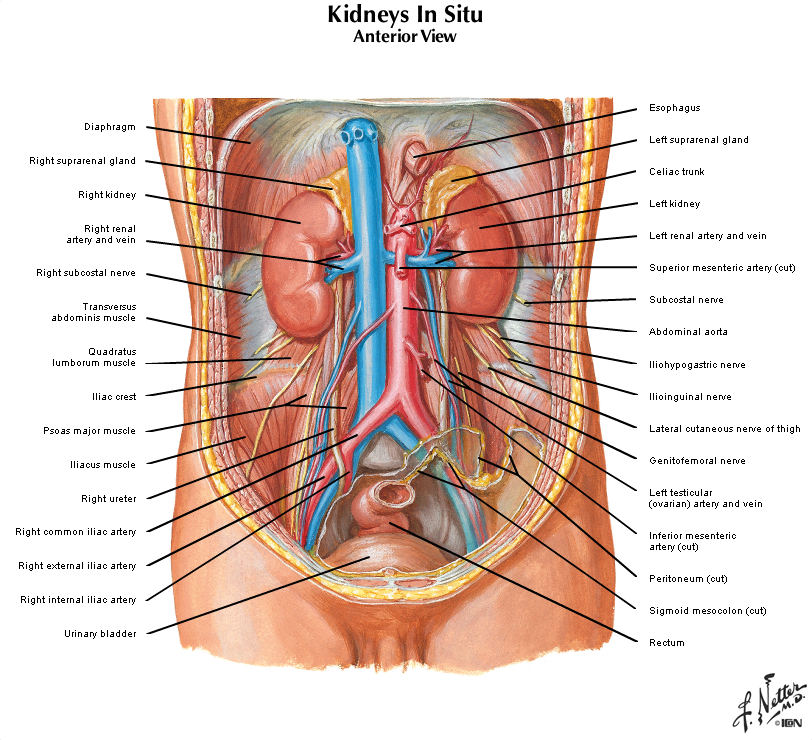
The kidney excretes soluble waste from the body and controls electrolyte balance. It consists of the cortex and the medulla. Within the outer cortex, glomeruli with delicate capillary loops are seen. The basement membrane is thin and without inflammation or thickening. Bowman’s capsule that surrounds the glomerulus is thin. The interstitium demonstrates no evidence of inflammation or fibrosis. In the areas between the glomeruli, tubules and arterioles are seen. The tubules are intact. The vessels exhibit no narrowing or wall thickening. The inner medulla of the kidney contains only tubules and blood vessels. Larger arteries and veins are located at the interface between cortex and medulla.
|
|
1-1. What is the differential diagnosis?
ANSWER
1-2. Which of the following genetic abnormalities is most likely to be the etiology this patient’s disease?
- Autosomal dominant mutation
- Autosomal recessive mutation
- Genomic imprinting
- Germline mosaicism
- Random X inactivation
ANSWER
1-3. Which of the following genes is most likely mutated in this patient?
- CFTR
- FBN1
- PAH
- PKD1
- PKHD1
ANSWER
1-4. This patient is at greatest risk for which of the following?
- Aortic stenosis
- Hepatic fibrosis
- Hypertension
- Portal hypertension
- Renal cell carcinoma
ANSWER
CASE NUMBER 7
(no virtual slide for this case)
Clinical History: A 48-year-old woman presents to the emergency department in labor. She has had no prenatal care. Delivery of the full-term infant is uncomplicated. An image of the neonate is provided.
Image Gallery:
7-1. What is the differential diagnosis?
ANSWER
Clinical History, continued (click here when instructed)
7-2. Which of the following processes best accounts for this patient’s karyotype?
- Amplification
- Chromosomal deletion
- Lyonization
- Mosaicism
- Premutation
ANSWER
7-3. Which of the following is the most likely etiology of this finding (ie. answer to question 2)?
- Amplification of the short arm of chromosome 21
- Excess trinucleotide repeats
- Meiotic nondisjunction
- Mitotic nondisjunction
ANSWER
7-4. Which of the following is most strongly associated with risk of having a child with this disease?
- Advanced maternal age
- In utero alcohol exposure
- Insufficient folate
- Oligohydramnios
- Toxoplasma infection
ANSWER
7-5. For which of the following is this patient at greatest risk?
- Alzheimer disease
- Hyperlipidemia
- Hypocalcemia
- Infertility
- Retinoblastoma
ANSWER
CASE NUMBER 6 - slide courtesy of UIndiana
[ImageScope] [WebScope]
Clinical History: A 2-year-old girl is brought to the pediatrician by her parents who noticed that she had a large right-sided abdominal mass. A CT scan was performed. Subsequently, the patient underwent a nephrectomy.
Image Gallery:
(Summary of Imaging Findings)
The CT scan shows a heterogeneous mass arising from the right kidney. A small sliver of residual kidney can be seen superomedial to the mass.
|
(Summary of Gross Findings)
Grossly, there is a solitary heterogeneous, lobulated, yellow-tan mass that distorts the kidney. The tumor appears well circumscribed. There are areas of hemorrhage and necrosis. Residual kidney can be seen at the top of the image.
|
(Summary of Microscopic Findings)
The slide shows a portion of normal kidney. The tumor displays triphasic histology: blastemal (small blue cells), stromal (spindle cells that are fibroblastic and often myxoid which is to say mucoid) and epithelial (glomerular structures and abortive tubules). Heterologous elements such as adipose tissue, cartilage and osteoid, are not seen here.
|
(Review Normal Histology)
Norm No. 2
[ImageScope] [WebScope]
The kidney excretes soluble waste from the body and controls electrolyte balance. It consists of the cortex and the medulla. Within the outer cortex, glomeruli with delicate capillary loops are seen. The basement membrane is thin and without inflammation or thickening. Bowman’s capsule that surrounds the glomerulus is thin. The interstitium demonstrates no evidence of inflammation or fibrosis. In the areas between the glomeruli, tubules and arterioles are seen. The tubules are intact. The vessels exhibit no narrowing or wall thickening. The inner medulla of the kidney contains only tubules and blood vessels. Larger arteries and veins are located at the interface between cortex and medulla.
|
|
6-1. What is the differential diagnosis?
ANSWER
6-2. Which of the following is most closely associated with poor prognosis in patients with this disease?
- Deletion of phenylalanine codon at position 508 (deltaF508)
- Diffuse anaplasia
- Intrauterine exposure to phenylalanine
- NMYC amplification
- X-linked inheritance
ANSWER
6-3. Which of the following genes is most commonly mutated in this tumor?
- CFTR
- FMR1
- IGF2
- RB
- WT1
ANSWER
6-4. Which of the following entities, associated with this tumor, is a disorder of genomic imprinting?
- Beckwith-Wiedemann syndrome
- Denys-Drash syndrome
- Fragile X syndrome
- Klinefelter syndrome
- WAGR (Wilms tumor, aniridia, genital abnormalities and mental retardation) syndrome
ANSWER
CASE NUMBER 539
[ImageScope] [WebScope]
Clinical History: A newborn baby is noted to have meconium ileus. His mother notices that when she kisses him, his skin tastes salty. A sweat chloride test was 110mEq/L (nl <60 mEq/L). Throughout childhood, he experiences persistent lung infections, chronic cough and obstructive pulmonary disease and, later in life, he develops exocrine pancreatic insufficiency with manifestations of malabsorption. He dies from cor pulmonale and an autopsy is performed.
Image Gallery:
(Summary of Gross Findings)
The bronchioles are extremely dilated and there are areas with extensive mucus plugging. The pulmonary parenchyma is consolidated by a combination of pneumonia and secretions.
|
(Summary of Microscopic Findings)
The bronchioles are dilated and filled with mucus admixed with abundant acute and chronic inflammatory cells. There is marked hyperplasia and hypertrophy of the mucus-secreting cells.
|
(Review Normal Histology)
Norm No. 24 Lung
[ImageScope] [WebScope]
The primary function of the lung is gas exchange. Therefore, alveoli have thin walls lined by thin flat pneumocytes and endothelial cells. There is no thickening or fibrosis of the interstitium. The bronchioli are lined with basally oriented ciliated columnar epithelium. The bronchi are lined by similar epithelium. There are mucous glands within the submucosa. The bronchial smooth muscle is not hypertrophied. The pulmonary vessels are patent with no evidence of intimal thickening or muscular hyperplasia.
|
|
539-1. Based on the clinical presentation what is the differential diagnosis?
ANSWER
539-2. Which of the following genes is most frequently mutated in this disease?
- APC
- CFTR
- COL3A1
- FMR1
- LDLR
ANSWER
539-3. Which of the following best accounts for the pulmonary findings in this patient?
- Decreased chloride secretion in respiratory epithelium
- High-volume surface fluid layer coating respiratory mucosa cells
- Hyperplasia of bronchial smooth muscle
- Hypoplasia of bronchiolar cartilage
- Increased water and sodium secretion in respiratory epithelium
ANSWER
539-4. Which of the following is most commonly found in patients with this disease?
- Cherry red macula
- Hepatosplenomegaly and bone lesions
- Increased plasma LDL
- Light colored hair and skin
- Pseudomonas aeruginosa infection
ANSWER
CASE NUMBER 601 (virtual slide in part 2 below)
Clinical History: A 44-year-old man has developed increasing arthritis pain, swelling of the feet, and reduced exercise tolerance over the past 3 years. He has smoked 1 pack of cigarettes per day for 20 years. Physical exam reveals metallic gray pigmentation of the sun-exposed skin on his face and bilateral swelling of the first and second metacarpophalangeal joints. His liver percussed to a span of 13 cm (normal 10.5 cm).
Laboratory studies include serum glucose of 201 mg/dL, creatinine 1.1 mg/dL and elevated serum iron of 175 mg/dL (normal, 50-150 mg/dL).
601-1. Which additional lab tests would you order?
- Transferrin saturation
- Chest X-ray
- MRI of the foot
- Joint aspirate
- Glucose tolerance test
ANSWER
--Clinical History, continued (click here when instructed)--
GENETIC DISORDERS Review Items
Key Vocabulary Terms (click here to search any additional terms on Stedman's Online Medical Dictionary)
LEARNING OBJECTIVES
Goal 1: Genetic Mechanisms of Developmental and Functional Abnormalities
Apply knowledge of the genetic mechanisms of disease to discuss how changes in the genome can cause developmental and functional abnormalities at the cellular, tissue and organism levels.
- Objective 1: Types of Mutations
Describe different types of mutations that can occur in human disease, and discuss how each of these can produce abnormalities in DNA transcription, and/or alterations in the type or amount of protein produced.
- Objective 2: Inheritance Patterns
Compare and contrast, with examples, the inheritance patterns of different types of Mendelian disorders.
- Objective 3: Genetic Diseases of Enzyme Function
Provide examples of genetic diseases associated with abnormal enzyme function, as opposed to those that produce abnormal structural or other non-enzyme proteins.
- Objective 4: Chromosomal Abnormalities
Discuss mechanisms that result in developmental abnormalities involving abnormal chromosomal number and provide examples of diseases associated with trisomies or chromosomal deletions.
- Objective 5: Multifactorial Inheritance and Environmental Factors
Discuss, with examples, disorders associated with multifactorial inheritance and describe how environmental factors can interact with genetic factors to produce disease.
- Objective 6: Non-classical Inheritance
Describe the pathophysiologic mechanisms that result in disorders of a non- classic inheritance and give clinical examples of each.
|