CASE NUMBER 221
(no virtual slide for this case)
Clinical History: A 62-year-old white man underwent total nephrectomy for renal cell carcinoma and subsequently received adjuvant chemotherapy. While on chemotherapy, he became leukopenic and thrombocytopenic. He presented to his oncologist with a 3-day history of a rash on his right shoulder.
Image Gallery:
(Summary of Gross Findings - click here)
There is a linear papulovesicular rash with lesions averaging 3-5 mm in size.
|
(Summary of Microscopic Findings - click here)
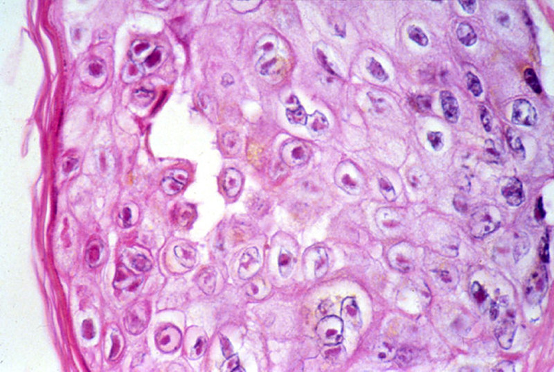
The epidermis shows spongiosis, vesicle formation, necrosis and ulceration. The epidermal cells have degrees of ballooning degeneration. Many intranuclear eosinophilic inclusion bodies are seen in these cells. Many keratinocytes within the vesicle also have multiple nuclei with nuclear "molding" and marginated chromatin. The upper dermis beneath a vesicle shows necrosis with little or no inflammatory reaction.
|
221-1. What is the differential diagnosis?
ANSWER
221-2. Which of the following is involved in the pathogenesis of this disease?
- IgA antibodies to gluten cross-react with reticulin
- Infection with Varicella-Zoster virus
- Keratinocyte destruction by cytotoxic T cells
- Linear deposits of IgG and complement at the dermoepidermal junction
- Mutation in the type VII collagen gene
ANSWER
221-3. Which of the following is a rare outcome that can be seen in this disease?
- B-cell lymphoma
- Facial hemiparalysis
- Myocarditis
- Toxic shock syndrome
- Vegetations on heart valves
ANSWER
CASE NUMBER 544
(no virtual slide for this case)
Clinical History: A 5-year-old boy is brought to the pediatrician because his parents notice that he walks on his toes and has difficulty climbing stairs. They state that he did not begin walking until he was 18 months old. Physical exam reveals bilaterally enlarged calves. A muscle biopsy is performed.
Image Gallery:
| |
(Summary of Microscopic Findings - click here)
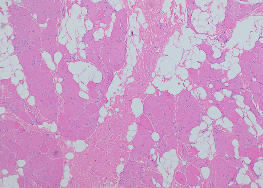
The low power view (left) shows fatty replacement of muscle fibers and hypertrophy and atrophy of fibers. The higher power view (right) shows marked fibrosis, fiber splitting, degeneration/myophagocytosis, and regeneration (internal nuclei).
|
544-1. What is the differential diagnosis?
ANSWER
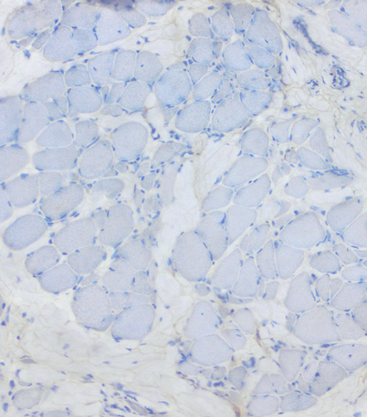
Immunohistochemical stains reveal complete absence of dystrophin protein and DNA sequencing confirms a deletion in the gene encoding this protein.
544-2. What is the correct diagnosis?
ANSWER
544-3. High serum levels of which of the following are frequently elevated in the early stages of this disease?
- Alkaline phosphatase
- Anti-Mi-2 antibodies
- Creatine kinase
- Dystrophia myotonica protein kinase
- Thyroid hormone
ANSWER
CASE NUMBER 523- slide courtesy of UMich
[ImageScope] [WebScope]
Clinical History: An 18-year-old man presents for his Army physical. The doctor notices multiple skin lesions distributed evenly across his body and irregular areas of hyperpigmentation. The patient states that he has had these lesions his entire life but that they had never bothered him. One of the lesions is biopsied.
Image Gallery:
(Summary of Clinical Findings - click here)
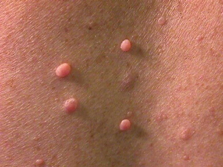
There are multiple soft, fleshy, pedunculated lesions present. In addition, there is a focus of hyperpigmentation consistent with a café au lait spot.
|
(Summary of Microscopic Findings - click here)
There is an infiltrative, non-circumscribed lesion present in the dermis that is composed of wavy or buckled spindle cells distributed amidst ropy collagen bundles and myxoid stroma. Mitotic activity, necrosis and cryptologic atypia are not seen.
|
Cerebrum
Slide 76 (cerebrum, luxol blue/cresyl violet) [WebScope] [ImageScope]
Slide 76b (toluidine blue & eosin) [WebScope] [ImageScope]
The cerebral cortex is loosely stratified into layers containing scattered nuclei of both neurons and glial cells. Examine the layered organization of the cerebral cortex using slide 76 stained with luxol blue/cresyl violet [ORIENTATION] (which stains white matter tracts and cell bodies) or toluidine blue and eosin [ORIENTATION] (TB&E, toluidine blue stains the nuclei and RER of cells whereas eosin stains membranes and axon tracts). Typically one or more sulci (infoldings) will extend inward from one edge of the section. Examine the gray matter on each side of the sulcus using first low and then high power. Neurons of the cerebral cortex are of varying shapes and sizes, but the most obvious are pyramidal cells. As the name implies, the cell body is shaped somewhat like a pyramid, with a large, branching dendrite extending from the apex of the pyramid toward the cortical surface, and with an axon extending downward from the base of the pyramid. In addition to pyramidal cells, other nuclei seen in these sections may belong to other neurons or to glial cells also present in the cortex. You may be able to see subtle differences in the distribution of cell types in rather loosely demarcated layers. There are 6 classically recognized layers of the cortex:
- Outer plexiform (molecular) layer: sparse neurons and glia
- Outer granular layer: small pyramidal and stellate neurons
- Outer pyramidal layer: moderate sized pyramidal neurons (should be able to see these in either luxol blue [example] or TB&E-stained [example] sections)
- Inner granular layer: densely packed stellate neurons (usually the numerous processes aren’t visible, but there are lots of nuclei reflecting the cell density)
- Ganglionic or inner pyramidal layer: large pyramidal neurons (should be able to see these in either luxol blue [example] or TB&E-stained [example] sections)
- Multiform cell layer: mixture of small pyramidal and stellate neurons
Pyramidal cells in layers III and V tend to be larger because their axons contribute to efferent projections that extend to other regions of the CNS –pyramidal neurons in layer V of motor cortices send projections all the way down to motor neurons in the spinal cord!
Deep to the gray matter of the cerebral cortex is the white matter that conveys myelinated fibers between different parts of the cortex and other regions of the CNS. Be sure you identify the white matter in both luxol blue [example] and TB&E-stained [example] sections, as it will appear differently in these two stains. Review the organization of gray and white matter in cerebral cortex vs. spinal cord.
|
523-1. What is the differential diagnosis?
ANSWER
523-2. A mutation in a gene encoding which of the following is most commonly mutated in patients with this disease?
- Dystrophin
- E-cadherin
- Merlin
- Neurofibromin
- Tuberin
ANSWER
523-3. Identification of which of the following is highly suggestive of this diagnosis?
- Hemangioblastoma
- Meningioma
- Plexiform neurofibroma
- Solitary cutaneous neurofibroma
- Vestibular schwannoma
ANSWER
Clinical History Continued: Ten years later, the patient presents to his primary care physician with a 3-week history of “burning” and “shooting” nerve pain in his right arm. MRI revealed a 5 cm mass within the brachial plexus. The mass was excised.
[ImageScope] [WebScope] - slide courtesy of UMich
(Summary of Microscopic Findings - click here)
Residual neurofibroma is present on the superior aspect of the tissue. The remaining tissue shows significant cytologic atypia, hypercellularity and mitotic figures.
|
|
Cerebrum
Slide 76 (cerebrum, luxol blue/cresyl violet) [WebScope] [ImageScope]
Slide 76b (toluidine blue & eosin) [WebScope] [ImageScope]
The cerebral cortex is loosely stratified into layers containing scattered nuclei of both neurons and glial cells. Examine the layered organization of the cerebral cortex using slide 76 stained with luxol blue/cresyl violet [ORIENTATION] (which stains white matter tracts and cell bodies) or toluidine blue and eosin [ORIENTATION] (TB&E, toluidine blue stains the nuclei and RER of cells whereas eosin stains membranes and axon tracts). Typically one or more sulci (infoldings) will extend inward from one edge of the section. Examine the gray matter on each side of the sulcus using first low and then high power. Neurons of the cerebral cortex are of varying shapes and sizes, but the most obvious are pyramidal cells. As the name implies, the cell body is shaped somewhat like a pyramid, with a large, branching dendrite extending from the apex of the pyramid toward the cortical surface, and with an axon extending downward from the base of the pyramid. In addition to pyramidal cells, other nuclei seen in these sections may belong to other neurons or to glial cells also present in the cortex. You may be able to see subtle differences in the distribution of cell types in rather loosely demarcated layers. There are 6 classically recognized layers of the cortex:
- Outer plexiform (molecular) layer: sparse neurons and glia
- Outer granular layer: small pyramidal and stellate neurons
- Outer pyramidal layer: moderate sized pyramidal neurons (should be able to see these in either luxol blue [example] or TB&E-stained [example] sections)
- Inner granular layer: densely packed stellate neurons (usually the numerous processes aren’t visible, but there are lots of nuclei reflecting the cell density)
- Ganglionic or inner pyramidal layer: large pyramidal neurons (should be able to see these in either luxol blue [example] or TB&E-stained [example] sections)
- Multiform cell layer: mixture of small pyramidal and stellate neurons
Pyramidal cells in layers III and V tend to be larger because their axons contribute to efferent projections that extend to other regions of the CNS –pyramidal neurons in layer V of motor cortices send projections all the way down to motor neurons in the spinal cord!
Deep to the gray matter of the cerebral cortex is the white matter that conveys myelinated fibers between different parts of the cortex and other regions of the CNS. Be sure you identify the white matter in both luxol blue [example] and TB&E-stained [example] sections, as it will appear differently in these two stains. Review the organization of gray and white matter in cerebral cortex vs. spinal cord.
|
523-4. What is the differential diagnosis?
ANSWER
523-5. Which of the following is the life time risk for developing this type of tumor in a patient with this disease?
- <1%
- 1 – 10%
- 21 - 30%
- 51 - 60%
- >90%
ANSWER
MUSCLE PATHOLOGY Review Items
Key Vocabulary Terms (click here to search any additional terms on Stedman's Online Medical Dictionary)
LEARNING OBJECTIVES
- Absolutely critical information you must know to practice medicine is in bold font.
- Important information that will be needed for routine patient care is in regular font.
- Information about less common diseases that you may encounter in clinical practice and that will probably appear on examinations is in italics
- Describe the structural features of normal skeletal muscle in terms of:
- gross morphology
- light microscopic appearance
- electron microscopic appearance
- histochemistry
- Describe proper skeletal muscle biopsy procedure, in terms of:
- choice of site
- biopsy technique
- techniques of fixation, processing, staining
- common artifacts seen
- limitations
- Describe the neuromuscular apparatus, and list disease processes and histopatholgic findings of diseases affecting the following components:
- Discuss the clinical approach and appropriate use of diagnostic tests in the evaluation of a patient with a myopathy.
- Describe the ways in which the following factors influence chemical injuries:
- Compare and contrast the clinical and pathologic features of skeletal muscle disorders:
- Compare and contrast the clinical and pathologic features of the following types of muscular dystrophy:
- Duchenne
- Becker
- Myotonic
- limb girdle
- Discuss the clinical and pathologic features of the following disorders:
- spinal muscular atrophy
- glycogenoses
- myasthenia gravis
- Lambert-Eaton myasthenic syndrome
- AIDS-associated myopathy
- viral myositis
- trichinosis
- cysticercosis
- polymyositis
EYE PATHOLOGY Review Items
Key Vocabulary Terms (click here to search any additional terms on Stedman's Online Medical Dictionary)
LEARNING OBJECTIVES
- Absolutely critical information you must know to practice medicine is in bold font.
- Important information that will be needed for routine patient care is in regular font.
- Information about less common diseases that you may encounter in clinical practice and that will probably appear on examinations is in italics
- Discuss the anatomy of the orbit and name the important compartments and tissues.
- Describe ocular findings in the following congenital conditions:
- trisomy 13
- trisomy 21
- congenital rubella
- congenital syphilis
- Discuss the clinical and pathologic features of inflammatory conditions of the eye and orbit:
|
|
|
|
|
|
|
|
|
- granulomatous inflammation
|
|
- sympathetic ophthalmia (uveitis)
|
- Compare and contrast the clinical and pathologic features of glaucoma:
- congenital
- primary angle-closure
- secondary angle closure
- open-angle
- Discuss the clinical and pathologic features of the following degenerative conditions:
- Discuss cataracts with regard to:
- associated diseases
- etiology
- classification
- morphology
- Discuss the clinical and pathologic features of the following diseases:
- retinopathy of prematurity (retrolental fibroplasia)
- retinitis pigmentosa
- macular degeneration
- retinal detachment
- Compare and contrast the clinical and pathologic features of he following vascular disorders:
- central retinal artery occlusion
- central retinal vein occlusion
- hypertensive retinopathy
- arteriosclerotic retinopathy
- diabetic retinopathy
- Describe the ocular lesions associated with:
- vitamin A deficiency
- methanol toxicity
- List the most frequent primary and metastatic malignancies of the:
- eyelid
- conjunctiva
- uvea (uveal tract)
- optic nerve
- Discuss the clinical and pathologic features of the following malignancies of the eye:
- malignant melanoma
- retinoblastoma
- metastatic malignancy
- Discuss the clinical and pathologic features of diseases of the optic nerve:
- papilledema
- optic neuritis
- optic atrophy
- Name the two most common causes of blindness in the world and the four most common causes of blindness in the United States
|