Case assignments by lab group and class day:
| |
TUESDAY |
THURSDAY |
| Labs 1, 2, & 3 |
|
|
| |
|
|
| Labs 4, 5, & 6 |
|
|
| |
|
|
CASE NUMBER 380
[ImageScope] [WebScope]
Clinical History:An 82-year-old man in a nursing home with a history of dementia developed left-sided upper and lower extremity weakness with hemiparesis of the left side of his face. He had been confined to his bed for the previous week due to a bad cold. A non-contrast CT showed no intraparenchymal hemorrhage but before tPA could be started, he became increasingly obtunded and was transferred to the ICU. He died the following afternoon. An autopsy was performed.
380-1. What is the differential diagnosis?
ANSWER
Image Gallery:
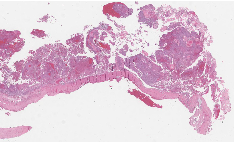
(Summary of Gross Findings)
At autopsy, a patent foramen ovale was noted. The right hemisphere of the brain was edematous. The cut sections showed moderate ventricular dilation. Blurring and discoloration of the gray-white junction was noted in the right frontoparietal regions along with parenchymal softening. A thrombus was found in the right MCA, occluding approximately 80% of the lumen.
|
(Summary of Microscopic Findings)
The sections demonstrate numerous red neurons with cytoplasmic eosinophilia, pyknotic nuclei, and perineuronal edema. Foci of parenchymal and perivascular hemorrhage are noted. In some areas a focal macrophage infiltrate is noted.
General Microscopic Features Observed with Injury to the CNS are as follows:
12 24 hours: acute neuronal injury manifests as red neurons24 hours to 2 weeks: subacute changes tissue necrosis, macrophage influx, reactive gliosis (nonspecific reactive change with increased astrocytes, microglia and oligodendrocytes)Repair after 2 weeks remove of necrotic tissue, gliosis
|
(Review CNS Histology)
Cerebrum
Slide 76 (cerebrum, luxol blue/cresyl violet) [WebScope] [ImageScope]
Slide 76b (toluidine blue & eosin) [WebScope] [ImageScope]
The cerebral cortex is loosely stratified into layers containing scattered nuclei of both neurons and glial cells. Examine the layered organization of the cerebral cortex using slide 76 stained with luxol blue/cresyl violet [ORIENTATION] (which stains white matter tracts and cell bodies) or toluidine blue and eosin [ORIENTATION] (TB&E, toluidine blue stains the nuclei and RER of cells whereas eosin stains membranes and axon tracts). Typically one or more sulci (infoldings) will extend inward from one edge of the section. Examine the gray matter on each side of the sulcus using first low and then high power. Neurons of the cerebral cortex are of varying shapes and sizes, but the most obvious are pyramidal cells. As the name implies, the cell body is shaped somewhat like a pyramid, with a large, branching dendrite extending from the apex of the pyramid toward the cortical surface, and with an axon extending downward from the base of the pyramid. In addition to pyramidal cells, other nuclei seen in these sections may belong to other neurons or to glial cells also present in the cortex. You may be able to see subtle differences in the distribution of cell types in rather loosely demarcated layers. There are 6 classically recognized layers of the cortex:
- Outer plexiform (molecular) layer: sparse neurons and glia
- Outer granular layer: small pyramidal and stellate neurons
- Outer pyramidal layer: moderate sized pyramidal neurons (should be able to see these in either luxol blue [example] or TB&E-stained [example] sections)
- Inner granular layer: densely packed stellate neurons (usually the numerous processes aren’t visible, but there are lots of nuclei reflecting the cell density)
- Ganglionic or inner pyramidal layer: large pyramidal neurons (should be able to see these in either luxol blue [example] or TB&E-stained [example] sections)
- Multiform cell layer: mixture of small pyramidal and stellate neurons
Pyramidal cells in layers III and V tend to be larger because their axons contribute to efferent projections that extend to other regions of the CNS –pyramidal neurons in layer V of motor cortices send projections all the way down to motor neurons in the spinal cord!
Deep to the gray matter of the cerebral cortex is the white matter that conveys myelinated fibers between different parts of the cortex and other regions of the CNS. Be sure you identify the white matter in both luxol blue [example] and TB&E-stained [example] sections, as it will appear differently in these two stains. Review the organization of gray and white matter in cerebral cortex vs. spinal cord.
|
|
380-2. List 5 risk factors for this disease process:
ANSWER
380-3. Which of the following arteries is most commonly occluded in this sort of patient?
- Anterior cerebral artery
- Anterior communicating artery
- Middle cerebral artery
- Ophthalmic artery
- Posterior cerebral artery
ANSWER
380-4. Which of the following is the proposed mechanism for hemorrhagic infarction?
- Acute vascular occlusion without resolution
- Direct trauma to brain parenchyma
- Global hypoxia due to hypotension
- Reperfusion of ischemic tissue
- Rupture of an aneurysm
ANSWER
380-5. What is the significance of the patent foramen ovale?
ANSWER
CASE NUMBER 427
[ImageScope] [WebScope]
Clinical History: A 78-year-old man presented to his primary care physician with a two-year history of headache that had worsened recently. In addition, he said that he had difficulty seeing. Visual field examination revealed bitemporal hemianopsia. No endocrine abnormalities were identified. He was admitted to the hospital for evaluation where MRI showed a 1.5 cm suprasellar mass. Screening CT showed no other abnormalities. While in the hospital, the patient contracted pneumonia and died. No other CNS pathology was noted at autopsy.
427-1. What is the differential diagnosis?
ANSWER
Image Gallery:
(Summary of Gross Findings)
The pituitary gland, weighing 3 gms and measuring 2 cms in greatest diameter, contained a large pink-gray soft tumor mass in the anterior lobe. The optic chiasm was slightly atrophic due to the compression of the tumor.
|
(Summary of Microscopic Findings)
The tumor occupies nearly the entire anterior lobe, so that only a thin rim of normal hypophyseal tissue is present in the subcapsular area. The tumor is composed of uniform cells arranged in a trabecular or sinusoidal pattern. The stroma is highly vascular. Some of the tumor cells contain fine eosinophilic granules, but most of them are chromophobes with special stains.
|
(Review Pituitary Histology)
Norm No. 14 Pituitary gland
[ImageScope] [WebScope]
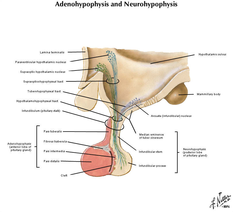
The pituitary is an endocrine gland with neurohypophysis and adenohypophysis. The neurohypophysis is composed of spindle cells that are derived from neural crest. These are neurosecretory cells. The adenohypophysis is an endocrine gland which is composed of three histological cell types, acidophils, basophils and chromophobes. The staining properties vary with the hormonal product. For example, growth hormone secreting cells are acidophils.
This section of brain is stained with hematoxylin and eosin. The tumor of the left side of the brain was due to the proliferation and infiltration of moderately pleomorphic fibrillary astrocytic cells. There is focal necrosis. Necrosis is the hallmark of glioblastoma. Better differentiated slower growing astrocytomas do not exhibit necrosis. |
|
427-2. Which of the following is true about this diagnosis?
- Classification is based on size and location
- It can be distinguished from hyperplasia by its pleomorphism and well-formed reticulin network
- G-protein mutations are commonly seen in somatotroph cell adenomas
- Gonadotroph adenomas cause Cushing syndrome
- Malignant transformation occurs in 25% of cases
ANSWER
CASE NUMBER 540 - slide courtesy of UMich
[ImageScope] [WebScope]
Clinical History: A 51-year-old woman presented to her GP with a 6-week history of increasingly severe headaches. A neurological exam was unremarkable. An MRI was performed and based on the presumptive diagnosis, an attempt at embolization of the mass was made. While she was in recovery, she experience a massive myocardial infarction and died. An autopsy was performed.
540-1. What is the differential diagnosis?
ANSWER
Image Gallery:
Imaging shows a dural based mass which is isointense to grey matter on both T1 and T2 weighted imaging, and demonstrate vivid contrast enhancement on both MRI and CT.
|
(Summary of Gross Findings)
There is a well circumscribed tumor attached to the dura. The tumor has compressed but has not invaded the brain.
|
(Summary of Microscopic Findings)
A dural based mass composed of bland cells with indistinct cell borders arranged in whorls. Rare psammoma bodies are present. Embolization material is seen in blood vessels.
|
(Review CNS Histology)
Cerebrum
Slide 76 (cerebrum, luxol blue/cresyl violet) [WebScope] [ImageScope]
Slide 76b (toluidine blue & eosin) [WebScope] [ImageScope]
The cerebral cortex is loosely stratified into layers containing scattered nuclei of both neurons and glial cells. Examine the layered organization of the cerebral cortex using slide 76 stained with luxol blue/cresyl violet [ORIENTATION] (which stains white matter tracts and cell bodies) or toluidine blue and eosin [ORIENTATION] (TB&E, toluidine blue stains the nuclei and RER of cells whereas eosin stains membranes and axon tracts). Typically one or more sulci (infoldings) will extend inward from one edge of the section. Examine the gray matter on each side of the sulcus using first low and then high power. Neurons of the cerebral cortex are of varying shapes and sizes, but the most obvious are pyramidal cells. As the name implies, the cell body is shaped somewhat like a pyramid, with a large, branching dendrite extending from the apex of the pyramid toward the cortical surface, and with an axon extending downward from the base of the pyramid. In addition to pyramidal cells, other nuclei seen in these sections may belong to other neurons or to glial cells also present in the cortex. You may be able to see subtle differences in the distribution of cell types in rather loosely demarcated layers. There are 6 classically recognized layers of the cortex:
- Outer plexiform (molecular) layer: sparse neurons and glia
- Outer granular layer: small pyramidal and stellate neurons
- Outer pyramidal layer: moderate sized pyramidal neurons (should be able to see these in either luxol blue [example] or TB&E-stained [example] sections)
- Inner granular layer: densely packed stellate neurons (usually the numerous processes aren’t visible, but there are lots of nuclei reflecting the cell density)
- Ganglionic or inner pyramidal layer: large pyramidal neurons (should be able to see these in either luxol blue [example] or TB&E-stained [example] sections)
- Multiform cell layer: mixture of small pyramidal and stellate neurons
Pyramidal cells in layers III and V tend to be larger because their axons contribute to efferent projections that extend to other regions of the CNS –pyramidal neurons in layer V of motor cortices send projections all the way down to motor neurons in the spinal cord!
Deep to the gray matter of the cerebral cortex is the white matter that conveys myelinated fibers between different parts of the cortex and other regions of the CNS. Be sure you identify the white matter in both luxol blue [example] and TB&E-stained [example] sections, as it will appear differently in these two stains. Review the organization of gray and white matter in cerebral cortex vs. spinal cord.
This section of brain is stained with hematoxylin and eosin. The tumor of the left side of the brain was due to the proliferation and infiltration of moderately pleomorphic fibrillary astrocytic cells. There is focal necrosis. Necrosis is the hallmark of glioblastoma. Better differentiated slower growing astrocytomas do not exhibit necrosis. |
540-2. Which of the following is the MOST COMMONLY mutated gene in patients with this tumor?
- IDHI
- MYC
- NF2
- TSC2
- VHL
ANSWER
540-3. Which of the following is a histologic feature frequently seen in this type of tumor?
- Brisk mitotic activity
- Cytoplasmic halo
- Homer Wright rosettes
- Necrosis
- Psammoma bodies
ANSWER
CASE NUMBER 541 - slide courtesy of UMich
[ImageScope] [WebScope]
Clinical History: A 34-year-old woman with no prior history of disease presents to the emergency department after her husband noticed her having a seizure. An MRI revealed a 3 x 4 x 4.2 cm mass in the right temporal lobe. A biopsy was performed.
541-1. What is the differential diagnosis?
ANSWER
Image Gallery:
The image on the left is a T2-weighted image and the one on the right is a T1-weighted image enhanced with gadolinium, a contrast agent that highlights brain tumors due to the breakdown of the blood-brain barrier. The image on the left shows peritumoral edema. In the image on the right, the central, dim area corresponds to areas of necrosis whereas the thickened ring shows pooling of gadolinium.
|
(Summary of Microscopic Findings)
Sections show areas of necrosis, hemorrhage and vascular proliferation. The tissue is hypercellular and displays atypical cells.
|
(Review CNS Histology)
Cerebrum
Slide 76 (cerebrum, luxol blue/cresyl violet) [WebScope] [ImageScope]
Slide 76b (toluidine blue & eosin) [WebScope] [ImageScope]
The cerebral cortex is loosely stratified into layers containing scattered nuclei of both neurons and glial cells. Examine the layered organization of the cerebral cortex using slide 76 stained with luxol blue/cresyl violet [ORIENTATION] (which stains white matter tracts and cell bodies) or toluidine blue and eosin [ORIENTATION] (TB&E, toluidine blue stains the nuclei and RER of cells whereas eosin stains membranes and axon tracts). Typically one or more sulci (infoldings) will extend inward from one edge of the section. Examine the gray matter on each side of the sulcus using first low and then high power. Neurons of the cerebral cortex are of varying shapes and sizes, but the most obvious are pyramidal cells. As the name implies, the cell body is shaped somewhat like a pyramid, with a large, branching dendrite extending from the apex of the pyramid toward the cortical surface, and with an axon extending downward from the base of the pyramid. In addition to pyramidal cells, other nuclei seen in these sections may belong to other neurons or to glial cells also present in the cortex. You may be able to see subtle differences in the distribution of cell types in rather loosely demarcated layers. There are 6 classically recognized layers of the cortex:
- Outer plexiform (molecular) layer: sparse neurons and glia
- Outer granular layer: small pyramidal and stellate neurons
- Outer pyramidal layer: moderate sized pyramidal neurons (should be able to see these in either luxol blue [example] or TB&E-stained [example] sections)
- Inner granular layer: densely packed stellate neurons (usually the numerous processes aren’t visible, but there are lots of nuclei reflecting the cell density)
- Ganglionic or inner pyramidal layer: large pyramidal neurons (should be able to see these in either luxol blue [example] or TB&E-stained [example] sections)
- Multiform cell layer: mixture of small pyramidal and stellate neurons
Pyramidal cells in layers III and V tend to be larger because their axons contribute to efferent projections that extend to other regions of the CNS –pyramidal neurons in layer V of motor cortices send projections all the way down to motor neurons in the spinal cord!
Deep to the gray matter of the cerebral cortex is the white matter that conveys myelinated fibers between different parts of the cortex and other regions of the CNS. Be sure you identify the white matter in both luxol blue [example] and TB&E-stained [example] sections, as it will appear differently in these two stains. Review the organization of gray and white matter in cerebral cortex vs. spinal cord.
This section of brain is stained with hematoxylin and eosin. The tumor of the left side of the brain was due to the proliferation and infiltration of moderately pleomorphic fibrillary astrocytic cells. There is focal necrosis. Necrosis is the hallmark of glioblastoma. Better differentiated slower growing astrocytomas do not exhibit necrosis. |
|
541-2. Which of the following genes is commonly mutated in this tumor?
- IDH2
- MYC
- TP53
- TSC
- VHL
ANSWER
541-3. Which of the following is true regarding this disease?
- In situ stages are detectable up to 5 years before diagnosis
- Median survival is less than two years
- Metastases to lung and liver are common
- Posterior fossa is the most common site of origin
- Psammoma bodies are commonly found histologically.
ANSWER
541-4. Which of the following is MOST LIKELY the cell of origin for this neoplasm?
- Astrocyte
- Ependymal cell
- Microglial cell
- Neuron
- Oligodendrocyte
ANSWER
CASE NUMBER 542 - slide courtesy of UMich
[ImageScope] [WebScope]
Clinical History: A 3-year-old boy was brought to the ED after an episode of projectile vomiting. According to his parents, 2 days earlier, he had begun vomiting after meals. Physical exam was notable for a temperature of 37.7C. His pupils were equal in size, but eye movements could not be fully evaluated due to his lack of cooperation. The ED physician believed he saw a down-beating nystagmus when the child opened his eyes. MRI shows the findings below. A biopsy was performed.
542-1. What is the differential diagnosis?
ANSWER
Image Gallery:
The first, T1-weighted, image shows a large, low signal mass along the midline of the posterior fossa with patchy contrast enhancement. The T2-weighted image shows a signal that is hyperintense compared to the gray matter; there is a focus of bright signal that may represent cyst formation. In both images, the tumor appears heterogeneous. The third image shows hydrocephalus which is due to obstruction.
|
(Summary of Microscopic Findings)
The lesion is composed of monomorphic small, round blue cells. They are primitive in appearance with fine chromatin and minute nucleoli. Necrosis is not present.
|
(Review CNS Histology)
Cerebrum
Slide 76 (cerebrum, luxol blue/cresyl violet) [WebScope] [ImageScope]
Slide 76b (toluidine blue & eosin) [WebScope] [ImageScope]
The cerebral cortex is loosely stratified into layers containing scattered nuclei of both neurons and glial cells. Examine the layered organization of the cerebral cortex using slide 76 stained with luxol blue/cresyl violet [ORIENTATION] (which stains white matter tracts and cell bodies) or toluidine blue and eosin [ORIENTATION] (TB&E, toluidine blue stains the nuclei and RER of cells whereas eosin stains membranes and axon tracts). Typically one or more sulci (infoldings) will extend inward from one edge of the section. Examine the gray matter on each side of the sulcus using first low and then high power. Neurons of the cerebral cortex are of varying shapes and sizes, but the most obvious are pyramidal cells. As the name implies, the cell body is shaped somewhat like a pyramid, with a large, branching dendrite extending from the apex of the pyramid toward the cortical surface, and with an axon extending downward from the base of the pyramid. In addition to pyramidal cells, other nuclei seen in these sections may belong to other neurons or to glial cells also present in the cortex. You may be able to see subtle differences in the distribution of cell types in rather loosely demarcated layers. There are 6 classically recognized layers of the cortex:
- Outer plexiform (molecular) layer: sparse neurons and glia
- Outer granular layer: small pyramidal and stellate neurons
- Outer pyramidal layer: moderate sized pyramidal neurons (should be able to see these in either luxol blue [example] or TB&E-stained [example] sections)
- Inner granular layer: densely packed stellate neurons (usually the numerous processes aren’t visible, but there are lots of nuclei reflecting the cell density)
- Ganglionic or inner pyramidal layer: large pyramidal neurons (should be able to see these in either luxol blue [example] or TB&E-stained [example] sections)
- Multiform cell layer: mixture of small pyramidal and stellate neurons
Pyramidal cells in layers III and V tend to be larger because their axons contribute to efferent projections that extend to other regions of the CNS –pyramidal neurons in layer V of motor cortices send projections all the way down to motor neurons in the spinal cord!
Deep to the gray matter of the cerebral cortex is the white matter that conveys myelinated fibers between different parts of the cortex and other regions of the CNS. Be sure you identify the white matter in both luxol blue [example] and TB&E-stained [example] sections, as it will appear differently in these two stains. Review the organization of gray and white matter in cerebral cortex vs. spinal cord.
This section of brain is stained with hematoxylin and eosin. The tumor of the left side of the brain was due to the proliferation and infiltration of moderately pleomorphic fibrillary astrocytic cells. There is focal necrosis. Necrosis is the hallmark of glioblastoma. Better differentiated slower growing astrocytomas do not exhibit necrosis. |
|
542-2. Which of the following is associated with a worse prognosis in patients with this disease?
- 1p/19 q deletions
- Epstein-Barr virus infection
- Mutations in the WNT signaling pathway
- MYC amplification
- t(8;21) translocation
ANSWER
542-3. Which of the following is true regarding this tumor?
- Approximately 25% of tumors arise in the cerebellum
- Median survival following treatment is 6 months
- Metastasis to soft tissue is common
- Radiation therapy is a useful adjunct
- This tumor accounts for approximately 80% of pediatric brain tumors
ANSWER
NERVOUS SYSTEM PATHOLOGY Review Items
Key Vocabulary Terms (click here to search any additional terms on Stedman's Online Medical Dictionary)
CNS-2: GOALS and LEARNING OBJECTIVES
Goal: Vascular Pathology of the Brain
Apply knowledge of the structure and function of the brain and general pathology concepts to discuss disorders resulting from altered blood supply and hypoxia to the brain.
- Objective 1: Stroke
Compare and contrast the two major mechanisms for stroke (ischemic vs hemorrhagic) and how treatment would differ for each.
- Objective 2: Cranial Hemorrhage
Compare and contrast the etiologies and clinical presentations of epidural, subdural, subarachnoid hemorrhages, basal ganglionic, and lobar hemorrhages.
- Objective 3: Hypertensive Hemorrhage
Describe the mechanism of hypertensive hemorrhage and name the common locations in which this occurs.
- Objective 4: Embolic Infarction
Describe how embolic infarcts differ from atherothrombotic infarcts in pathologic appearance and name three sources of emboli.
- Objective 5: Acute vs Chronic Brain Infarction
Compare and contrast the gross and histopathologic appearance of acute versus remote brain infarction.
Goal: Congenital Malformations
Apply knowledge of the clinical, anatomic and neuropathologic principles to describe the clinicopathologic features and consequences of congenital malformations.
- Objective 1: Congenital Malformations
Name the most common forms of congenital malformations of the CNS and outline their clinical presentation, natural history, and long- and short-term complications.
Goal: Perinatal Brain Injury
Apply knowledge of the clinical, anatomic and neuropathologic principles to describe the clinicopathologic features and consequences of perinatal brain injury.
- Objective 1: Perinatal Brain Injury
Describe the clinicopathologic features of perinatal brain injury and outline the clinical presentation, natural history, and long- and short-term complications
Goal: CNS Neoplasia
Apply knowledge of the pathological and molecular basis of common brain tumors to describe their clinical behavior, effects on the nervous system, and therapies.
- Objective 1: Features of Brain Tumors
Explain the pathophysiology underlying the signs and symptoms associated with brain tumors.
- Objective 2: Classification of Brain Tumors
List the common types of brain tumors that affect the cerebrum, the cerebellum, the meninges, and the cranial nerves in adults and children; and outline their molecular basis and clinicopathologic features.
- Objective 3: Hereditary Tumor Syndromes
Describe the major hereditary tumor syndromes of the nervous system, the genes responsible for each syndrome, and the spectrum of tumors associated with each syndrome.
- Objective 4: Grading of Brain Tumors
Explain the pathophysiologic basis for grading primary brain tumors and discuss how grading relates to prognosis and governs patient management.
- Objective 5: Carcinomas Metastasizing to the CNS
List carcinomas that commonly metastasize to the central nervous system and describe the locations in which metastases may be seen.
|