CASE NUMBER 503
(no virtual slide for this case)
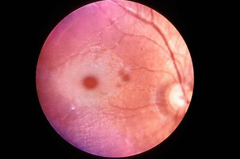
Clinical History: A mother brought her 3-month-old infant to the pediatrician because the child had been alternately crying inconsolably or lying down listlessly; vomiting for the last 3 days, and had a mild fever. Physical exam reveals cherry red maculas. CSF analysis shows elevated protein levels. Gene sequencing reveals a mutation in the a-subunit of hexosaminidase A.
Image Gallery:
503-1. What is the differential diagnosis?
ANSWER
503-2. This disease is classified as which of the following?
- Glycogenosis
- Mucopolylipidosis
- Mucopolysaccharidosis
- Sphingolipidosis
- Sulfatidosis
ANSWER
503-3. Which of the following is true regarding this disease?
- Boys are affected more commonly than girls
- Most patients recover full neurological function in their teens
- The “cherry red macula” is due to retinal hemangiomas
- There is an increased incidence in Ashknazi Jews
- These patients are at increased risk for colon cancer
ANSWER
CASE NUMBER 427
[ImageScope] [WebScope]
Clinical History: A 78-year-old man presented to his primary care physician with a two-year history of headache that had worsened recently. In addition, he said that he had difficulty seeing. Visual field examination revealed bitemporal hemianopsia. No endocrine abnormalities were identified. He was admitted to the hospital for evaluation where MRI showed a 1.5 cm suprasellar mass. Screening CT showed no other abnormalities. While in the hospital, the patient contracted pneumonia and died. No other CNS pathology was noted at autopsy.
Image Gallery:
(Summary of Gross Findings - click here)
The pituitary gland, weighing 3 gms and measuring 2 cms in greatest diameter, contained a large pink-gray soft tumor mass in the anterior lobe. The optic chiasm was slightly atrophic due to the compression of the tumor.
|
(Summary of Microscopic Findings - click here)
The tumor occupies nearly the entire anterior lobe, so that only a thin rim of normal hypophyseal tissue is present in the subcapsular area. The tumor is composed of uniform cells arranged in a trabecular or sinusoidal pattern. The stroma is highly vascular. Some of the tumor cells contain fine eosinophilic granules, but most of them are chromophobes with special stains.
|
(Review Normal Histology - click here)
Cerebrum
Slide 76 (cerebrum, luxol blue/cresyl violet) [WebScope] [ImageScope]
Slide 76b (toluidine blue & eosin) [WebScope] [ImageScope]
The cerebral cortex is loosely stratified into layers containing scattered nuclei of both neurons and glial cells. Examine the layered organization of the cerebral cortex using slide 76 stained with luxol blue/cresyl violet [ORIENTATION] (which stains white matter tracts and cell bodies) or toluidine blue and eosin [ORIENTATION] (TB&E, toluidine blue stains the nuclei and RER of cells whereas eosin stains membranes and axon tracts). Typically one or more sulci (infoldings) will extend inward from one edge of the section. Examine the gray matter on each side of the sulcus using first low and then high power. Neurons of the cerebral cortex are of varying shapes and sizes, but the most obvious are pyramidal cells. As the name implies, the cell body is shaped somewhat like a pyramid, with a large, branching dendrite extending from the apex of the pyramid toward the cortical surface, and with an axon extending downward from the base of the pyramid. In addition to pyramidal cells, other nuclei seen in these sections may belong to other neurons or to glial cells also present in the cortex. You may be able to see subtle differences in the distribution of cell types in rather loosely demarcated layers. There are 6 classically recognized layers of the cortex:
- Outer plexiform (molecular) layer: sparse neurons and glia
- Outer granular layer: small pyramidal and stellate neurons
- Outer pyramidal layer: moderate sized pyramidal neurons (should be able to see these in either luxol blue [example] or TB&E-stained [example] sections)
- Inner granular layer: densely packed stellate neurons (usually the numerous processes aren’t visible, but there are lots of nuclei reflecting the cell density)
- Ganglionic or inner pyramidal layer: large pyramidal neurons (should be able to see these in either luxol blue [example] or TB&E-stained [example] sections)
- Multiform cell layer: mixture of small pyramidal and stellate neurons
Pyramidal cells in layers III and V tend to be larger because their axons contribute to efferent projections that extend to other regions of the CNS –pyramidal neurons in layer V of motor cortices send projections all the way down to motor neurons in the spinal cord!
Deep to the gray matter of the cerebral cortex is the white matter that conveys myelinated fibers between different parts of the cortex and other regions of the CNS. Be sure you identify the white matter in both luxol blue [example] and TB&E-stained [example] sections, as it will appear differently in these two stains. Review the organization of gray and white matter in cerebral cortex vs. spinal cord.
This section of brain is stained with hematoxylin and eosin. The tumor of the left side of the brain was due to the proliferation and infiltration of moderately pleomorphic fibrillary astrocytic cells. There is focal necrosis. Necrosis is the hallmark of glioblastoma. Better differentiated slower growing astrocytomas do not exhibit necrosis. |
427-1. What is the differential diagnosis?
ANSWER
427-2. Which of the following is true about this diagnosis?
- Classification is based on size and location
- It can be distinguished from hyperplasia by its pleomorphism and well-formed reticulin network
- G-protein mutations are commonly seen in somatotroph cell adenomas
- Gonadotroph adenomas cause Cushing syndrome
- Malignant transformation occurs in 25% of cases
ANSWER
CASE NUMBER 540 - slide courtesy of UMich
[ImageScope] [WebScope]
Clinical History: A 51-year-old woman presented to her GP with a 6-week history of increasingly severe headaches. A neurological exam was unremarkable. An MRI was performed. When the patient left her doctor’s office, she was hit by a car and died. An autopsy was performed.
Imaging: Imaging shows a dural based mass which is isointense to grey matter on both T1 and T2 weighted imaging, and demonstrate vivid contrast enhancement on both MRI and CT.
Image Gallery:
540-1. What is the differential diagnosis?
ANSWER
540-2. Which of the following is the most commonly mutated gene in patients with this tumor?
- IDHI
- MYC
- NF2
- TSC2
- VHL
ANSWER
540-3. Which of the following is a histologic feature frequently seen in this type of tumor?
- Brisk mitotic activity
- Cytoplasmic halo
- Homer Wright rosettes
- Necrosis
- Psammoma bodies
ANSWER
CASE NUMBER 541 - slide courtesy of UMich
[ImageScope] [WebScope]
Clinical History: A 34-year-old woman with no prior history of disease presents to the emergency department after her husband noticed her having a seizure. An MRI revealed a 3 x 4 x 4.2 cm mass in the right temporal lobe. A biopsy was performed.
Image Gallery:
(Summary of Radiologic Findings - click here)
The image on the left is a T2-weighted image and the one on the right is a T1-weighted image enhanced with gadolinium, a contrast agent that highlights brain tumors due to the breakdown of the blood-brain barrier. The image on the left shows peritumoral edema. In the image on the right, the central, dim area corresponds to areas of necrosis whereas the thickened ring shows pooling of gadolinium.
|
(Summary of Microscopic Findings - click here)
Sections show areas of necrosis, hemorrhage and vascular proliferation. The tissue is hypercellular and displays atypical cells. Foamy histiocytes with hemosiderin pigment are present. Look for palisading I didnt see any, but I may have missed it.
|
|
541-1. What is the differential diagnosis?
ANSWER
541-2. Which of the following genes is commonly mutated in this tumor?
- IDH2
- MYC
- P53
- TSC
- VHL
ANSWER
541-3. Which of the following is true regarding this disease?
- In situ stages are detectable up to 5 years before diagnosis
- Median survival is less than two years
- Metastases to lung and liver are common
- Posterior fossa is the most common site of origin
- Psammoma bodies are commonly found histologically.
ANSWER
541-4. Which of the following is most likely the cell of origin for this neoplasm?
- Astrocyte
- Ependymal cell
- Microglial cell
- Neuron
- Oligodendrocyte
ANSWER
CASE NUMBER 542 - slide courtesy of UMich
[ImageScope] [WebScope]
Clinical History: A 3-year-old boy was brought to the ED after an episode of projectile vomiting. According to his parents, 2 days earlier, he had begun vomiting after meals. Physical exam was notable for a temperature of 37.7C. His pupils were equal in size, but eye movements could not be fully evaluated due to his lack of cooperation. The ED physician believed he saw a down-beating nystagmus when the child opened his eyes. MRI shows the findings below. A biopsy was performed.
Image Gallery:
(Summary of Imaging Studies - click here)
The first, T1-weighted, image shows a large, low signal mass along the midline of the posterior fossa with patchy contrast enhancement. The T2-weighted image shows a signal that is hyperintense compared to the gray matter; there is a focus of bright signal that may represent cyst formation. In both images, the tumor appears heterogeneous. The third image shows hydrocephalus which is due to obstruction.
|
(Summary of Microscopic Findings - click here)
The lesion is composed of monomorphic small, round blue cells. They are primitive in appearance with fine chromatin and minute nucleoli. Necrosis is not present. Homer Wright rosettes are present focally, but you need to look for them.
|
|
542-1. What is the differential diagnosis?
ANSWER
542-2. Which of the following is associated with a worse prognosis in patients with this disease?
- 1p/19 q deletions
- Epstein-Barr virus infection
- Mutations in the WNT signaling pathway
- MYC amplification
- t(8;21) translocation
ANSWER
542-3. Which of the following is true regarding this tumor?
- Approximately 25% of tumors arise in the cerebellum
- Median survival following treatment is 6 months
- Metastasis to soft tissue is common
- Radiation therapy is a useful adjunct
- This tumor accounts for approximately 80%% of pediatric brain tumors
ANSWER
NERVOUS SYSTEM PATHOLOGY Review Items
Key Vocabulary Terms (click here to search any additional terms on Stedman's Online Medical Dictionary)
LEARNING OBJECTIVES
- Absolutely critical information you must know to practice medicine is in bold font.
- Important information that will be needed for routine patient care is in regular font.
- Information about less common diseases that you may encounter in clinical practice and that will probably appear on examinations is in italics
- Describe the morphology and function of the following CNS cells:
|
|
|
- choroid plexus epithelial cells
|
|
|
|
|
- Compare CNS myelin with PNS myelin, in terms of:
- cells of elaboration
- structure and function
- reactions to injury and destruction
- regenerative potential
- Discuss normal CSF in terms of:
- sites of formation
- circulation patterns
- sites of absorption
- pressure
- glucose and protein levels
- cell types present
- Describe the blood-brain barrier (BBB) in terms of:
- physiologic definition
- anatomic counterparts
- morphologic alterations
- areas of absence
- Describe the morphology and function of the following CNS cells:
|
|
|
|
|
|
- ischemic neuronal necrosis
|
|
|
|
|
|
- Compare and contrast the following types of cerebral edema and their significance:
- cytotoxic
- vasogenic
- interstitial
- Compare and contrast the clinical findings and sequelae of herniation of the brain:
- subfalcine (cingulate gyrus)
- transtentorial (uncal)
- foraminal (tonsillar)
- Correlate destructive lesions in specific areas of the CNS with corresponding functional consequences.
- Compare and contrast:
- communicating hydrocephalus
- non-communicating hydrocephalus
- hydrocephalus ex vacuo
- Describe the following congenital abnormalities and their clinical phenotype:
|
- spina bifida/meningomyelocele
|
- Chiari type I malformation
|
|
- Chiari type II (Arnold-Chiari) malformation
|
|
- Dandy-Walker malformation
|
|
|
- agenesis of corpus callosum
|
|
|
|
|
- Compare and contrast genetics, clinical presentation and pathology of inborn errors of metabolism:
|
- spina bifida/meningomyelocele
|
- Chiari type I malformation
|
|
- Chiari type II (Arnold-Chiari) malformation
|
|
- Dandy-Walker malformation
|
|
|
- agenesis of corpus callosum
|
|
|
|
|
- Describe the effects of hypoxia/ischemia on the late gestational/perinatal brain, including the pathophysiologic mechanisms underlying the following:
|
|
- germinal matrix hemorrhage
|
|
- periventricular leukomalacia
|
|
- Discuss the clinical and pathologic features of the following processes:
- Compare and contrast the clinical and pathologic features of CNS aneurysms:
- saccular ("berry"
- atherosclerotic
- Charcot-Bouchard
- mycotic
- Compare and contrast the clinical and pathologic features of CNS vascular malformations:
- arteriovenous malformation
- cavernous angioma
- capillary telangiectasia
- List the ways in which hypertension may harm the brain.
- Compare and contrast the clinical and pathologic features of:
- hypertensive encephalopathy
- hypoxic encephalopathy
- multi infarct dementia
- Compare and contrast the clinical and pathologic features of CNS infarcts:
- nonhemorrhagic (pale, anemic)
- hemorrhagic (red)
- border zone (watershed)
- incomplete
- spinal cord
- Compare and contrast clinical presentations of infarcts in these vascular territories:
- middle cerebral
- vertebrobasilar
- internal carotid
- Describe the interrelationship between hypotension and watershed infarcts.
- Explain the basis of the reperfusion theory of causation of hemorrhagic cerebral infarcts.
- Compare and contrast the clinical and pathologic features:
- skull fracture
- parencymal brain injury
- vascular brain injury
- Compare and contrast open vs. closed head injury, complications and prognosis.
- Compare and contrast the clinical and pathologic features of the following entities:
- pyogenic meningitis
- tuberculous/mycobacterial meningoencephalitis
- viral meningoencephalitis
- fungal meningitis
- neurosyphilis
- neuroborreliosis (Lyme disease)
- rickettsial infection
- protozoal infection
- List the common bacterial agents of acute pyogenic meningitis, and the age group that each most frequently affects.
- Compare and contrast the clinical and pathologic features:
- brain abscess
- subdural empyema
- extradural abscess
- Compare and contrast the clinical and pathologic features of viral meningoencephalitis:
- arboviral encephalitides
- herpes simplex viral encephalitis
- varicella-zoster viral encephalitis
- cytomegalovirus (CMV) encephalitis
- poliomyelitis
- rabies
- human immunodeficiency virus (HIV) infections
- HIV meningoencephalitis (subacute encephalitis)
- vacuolar myelopathy
- progressive multifocal leukoencephalopathy (PML)
- subacute sclerosing panencephalitis (SSPE)
- Discuss the clinical and pathologic features of the following prion diseases:
- Creutzfeldt-Jakob disease (CJD)
- variant CJD (vCJD, "mad cow" disease)
- kuru
- scrapie
- Compare and contrast the clinical and pathologic features degenerative diseases:
|
- olivopontocerebellar atrophy
|
|
|
|
- spinocerebellar degeneration
|
- progressive supranuclear palsy
|
- amyotrophic lateral sclerosis (ALS)
|
- corticobasal degeneration
|
|
- striatonigral degeneration
|
|
|
|
- Describe multiple sclerosis (MS) in terms of:
- geographic distribution
- etiology
- age at onset
- distribution of lesions
- morphology
- clinical course
- Discuss the following nervous system disorders:
|
- carbon monoxide poisoning
|
- acute ethanol intoxication
|
|
|
- central pontine myelinolysis (CPM)
|
|
|
- Discuss the clinical and pathologic features of the following nutritional disorders:
- Wernicke encephalopathy
- Korsakoff psychosis
- neuropathic beriberi
- subacute combined degeneration
- Explain the concepts of benign vs. malignant neoplasms of the CNS.
- Compare and contrast the clinical, pathologic, epidemiologic and genetic features of the following CNS neoplasms:
- colloid cyst of third ventricle
|
|
|
|
|
|
|
|
|
|
|
|
|
|
|
|
|
|
|
|
|
|
|
- malignant peripheral nerve sheath tumor
|
|
|
- Compare and contrast the clinical, pathologic and genetic features of the following phakomatoses:
|
|
|
|
|
- von Hippel-Lindau syndrome
|
- Discuss the clinical and pathologic features of the following disorders of the PNS:
|
|
|
|
|
- paraproteinemia-associated neuropathy
|
|
|
- AIDS-associated peripheral neuropathy
|
|
- hereditary motor & sensory neuropathy (HMSN)
|
|
- type I [Charcot-Marie-Tooth disease (CMT) 1]
|
|
- type III (Dejerine-Sottas disease)
|
|
|