CASE NUMBER 452
[ImageScope] [WebScope]
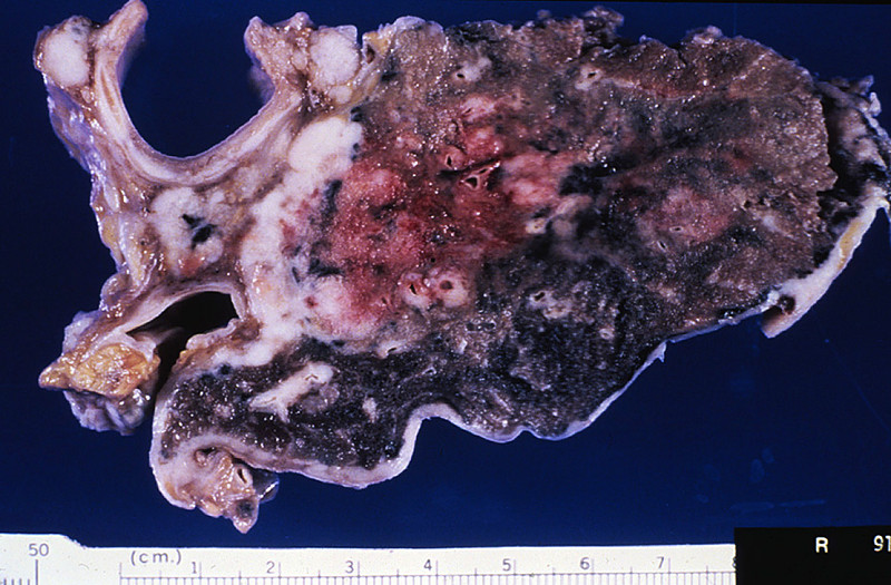
Clinical History: A 45-year-old man presented to his primary care physician with a 6-week history of non-productive cough. Over the last month or so, he had lost 15 pounds and occasionally experienced hemoptysis. He was originally from India, but moved to the United States nine years ago. Physical exam revealed harsh breath sounds with crackling rales over the right posterior lung base. Samples were taken for culture analysis; however, as he was leaving the doctor’s office, he experienced severe coughing with massive hemoptysis and died.
Image Gallery:
(Summary of Gross and Lab Findings - click here)
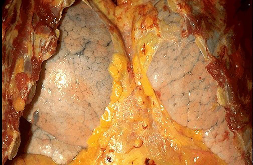
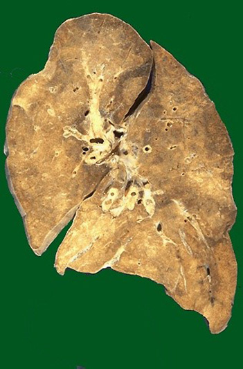
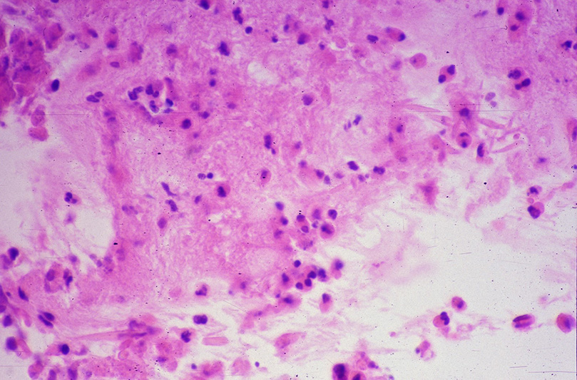
The lungs were heavy and had fibrous pleural adhesions. A large cavity containing caseous material was found in the apex of the left lung. The cavity communicated with a bronchus. In the left lower lobe there was large area of consolidation, which exuded creamy, yellow-gray, caseous material from the cut surface.
|
(Summary of Microscopic Findings - click here)
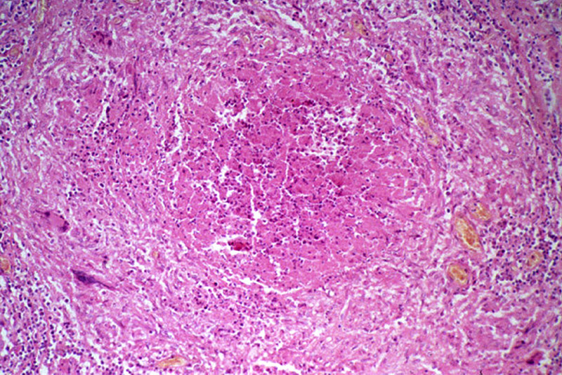
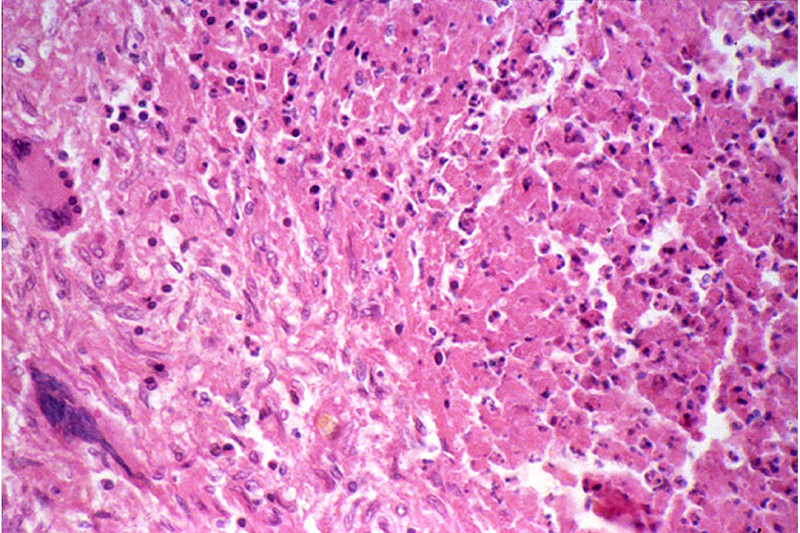
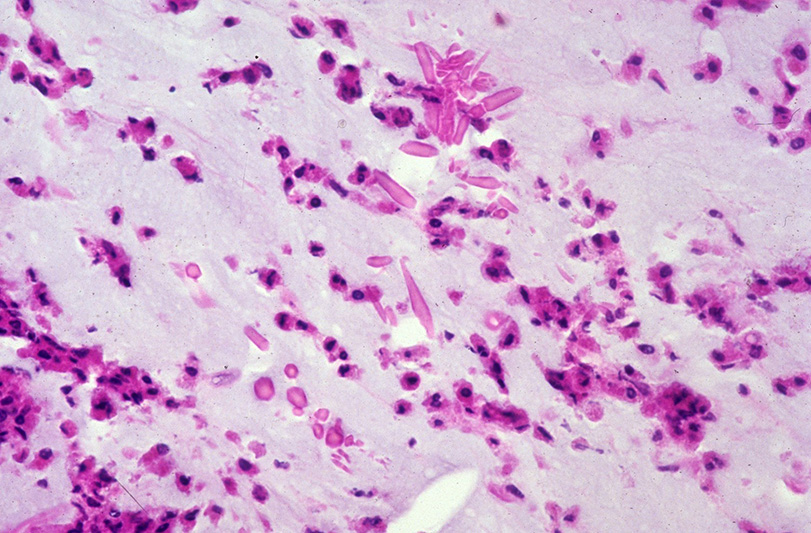
This section shows many foci of caseous necrosis, which is associated with marked proliferation of histiocytes around the caseous foci.
|
(Review Normal Histology - click here)
Norm No. 24 Lung
[ImageScope] [WebScope]
The primary function of the lung is gas exchange. Therefore, alveoli have thin walls lined by thin flat pneumocytes and endothelial cells. There is no thickening or fibrosis of the interstitium. The bronchioli are lined with basally oriented ciliated columnar epithelium. The bronchi are lined by similar epithelium. There are mucous glands within the submucosa. The bronchial smooth muscle is not hypertrophied. The pulmonary vessels are patent with no evidence of intimal thickening or muscular hyperplasia.
|
452-1. Which of the following is the most likely diagnosis?
- Berylliosis
- Leprosy
- Sarcoidosis
- Tuberculosis
- Viral pneumonia
ANSWER
452-2. Which of the following cells does Mycobacterium tuberculosis typically infect?
- B cells
- Cytotoxic T cells
- Macrophages
- Natural killer cells
- T helper cells
ANSWER
452-3. Immunity to M. tuberculosis is primarily mediated by which of the following?
- Plasma cells
- Natural killer cells
- TH1 cells
- TH2 cells
- TH17 cells
ANSWER
CASE NUMBER 602
(no virtual slides for this case)
Clinical History: History is obtained from paramedics and from the patient's boyfriend. 41 year old woman with a history of severe asthma and reactive airways disease. This began in childhood, and attacks have necessitated intubation on a number of previous occasions. She was at a pharmacy and began to have an attack. Per her boyfriend, she began prompt self-treatment with a portable nebulizer, and EMS was called. By the time paramedics arrived, she was in severe respiratory distress. Intramuscular epinephrine was administered but she suffered a cardiac arrest. Upon arrival at the Emergency Department, she was pulseless and apneic. She was intubated and 40 minutes of CPR were administered, but a pulse was never re-established. She was pronounced dead.
Image Gallery:
602-1. Which of the following is the most likely diagnosis?
- Bronchiectasis
- Aspiration
- Bronchial asthma
- Centrilobular emphysema
- Chronic bronchitis
- Obstructive sleep apnea
ANSWER
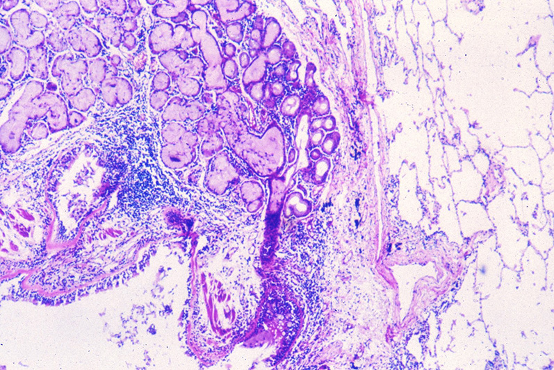
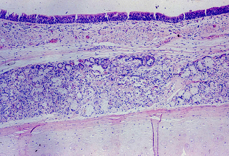
602-2. At autopsy, which of the following is the most likely histologic finding in the lungs?
- Bronchial smooth muscle hypertrophy with proliferation of eosinophils
- Diffuse alveolar damage with leakage of protein-rich fluid into alveolar spaces
- Dilation of air spaces with destruction of alveolar walls
- Hyperplasia of bronchial mucus-secreting submucosal glands
- Permanent bronchial dilation caused by chronic infection, with bronchi filled with mucus and neutrophils
ANSWER
CASE NUMBER 603
(no virtual slides for this case)
Clinical History: A 62-year-old man is a smoker with a 10-year history of cough productive of copious mucopurulent sputum. Over the past 6 months, he has developed progressive dyspnea. Physical examination shows bilateral pedal edema and a soft but enlarged liver. A chest radiograph shows bilateral pleural effusions and a prominent heart border on the right side. Arterial blood gas determinations show PO2 of 60 mm Hg, PCO2 55 mm Hg, pH 7.31, and HCO3- 28 mEq/L. The patient is intubated and placed on a ventilator, and he requires increasing amounts of oxygen.
Image Gallery:
603-1. Which of the following is the most likely diagnosis?
- Bronchiectasis
- Aspiration
- Bronchial asthma
- Centrilobular emphysema
- Chronic bronchitis
- Obstructive sleep apnea
ANSWER
603-2. At autopsy, which of the following microscopic findings is most likely to be characteristic of his underlying pulmonary disease?
- Infiltrates of eosinophils
- Extensive interstitial fibrosis
- Granulomas in bronchovascular distribution
- Carcinoma filling lymphatic spaces
- Hypertrophy of bronchial submucosal glands
ANSWER
CASE NUMBER 560
[WebScope] [ImageScope]
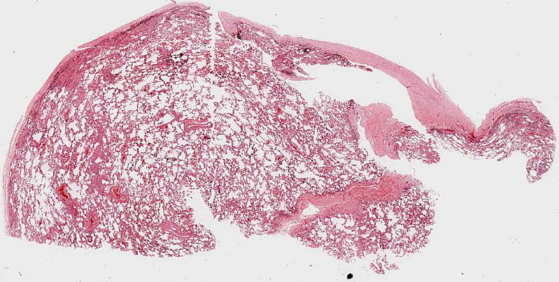
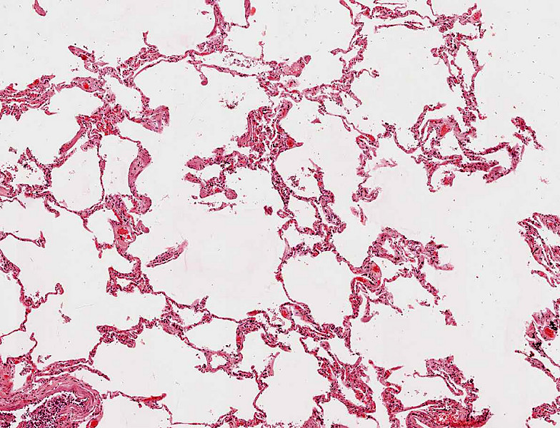
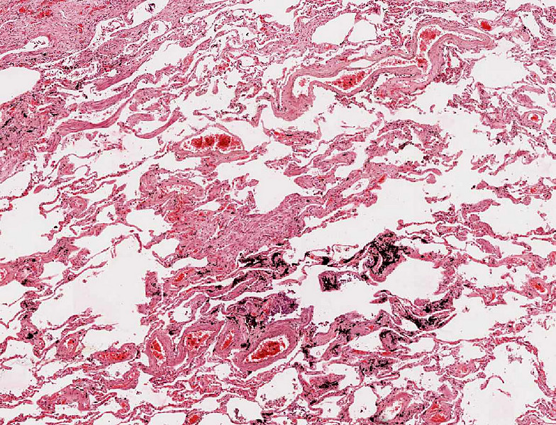
Clinical History: A 72-year-old man was found dead in his home by his wife. She told the medical examiner that he was a retired shipyard worker who had smoked 1 pack of cigarettes/day since he was 19. Autopsy revealed that the cause of death was myocardial infarction.
Image Gallery (slide specimen and images courtesy of the University of Michigan):
560-1. Which of the following is the most likely diagnosis?
- Adenocarcinoma
- Histoplasmosis
- Asbestosis
- Mesothelioma
- Silicosis
ANSWER
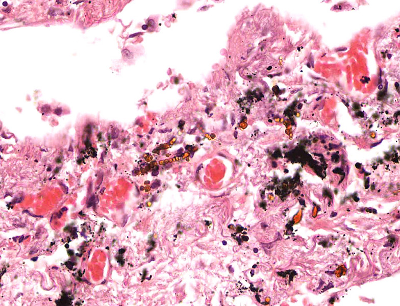
560-2. What are the golden-brown objects seen in the microscopic images?
- Beryllium fragments
- Charcot-Leyden crystals
- Ferruginous bodies
- Fungal spores
- Hyaline membranes
ANSWER
560-3. Patients with this disease are at greatest risk for developing which of the following complications?
- Acute interstitial pneumonia
- Bronchogenic carcinoma
- Diffuse alveolar damage
- Hypersensitivity pneumonitis
- Sarcoidosis
ANSWER
560-4. Which of the following pulmonary function test results is consistent with this patient’s diagnosis?
- Decreased forced vital capacity
- Decreased maximal expiratory flow rate
- Increased forced expiratory volume at 1 second
- Increased residual volume
- Increased total lung capacity
ANSWER
CASE NUMBER 126
[ImageScope] [WebScope]
Clinical History: A 50-year-old man presented to his primary care physician with a 2-month history of increasing dyspnea and unexplained weight loss. Chest X-ray revealed a 5 cm right upper lobe mass. While he was waiting in radiology for a staging CT, he suddenly collapsed. He could not be resuscitated.
Image Gallery:
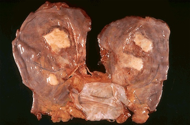
(Summary of Gross and Lab Findings - click here)
The lungs were remarkable for a 5 cm mass in the right upper lobe. The cut surfaces were friable and yellowish-gray. Regional lymph nodes were filled with similar necrotic tumor.
|
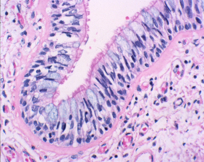
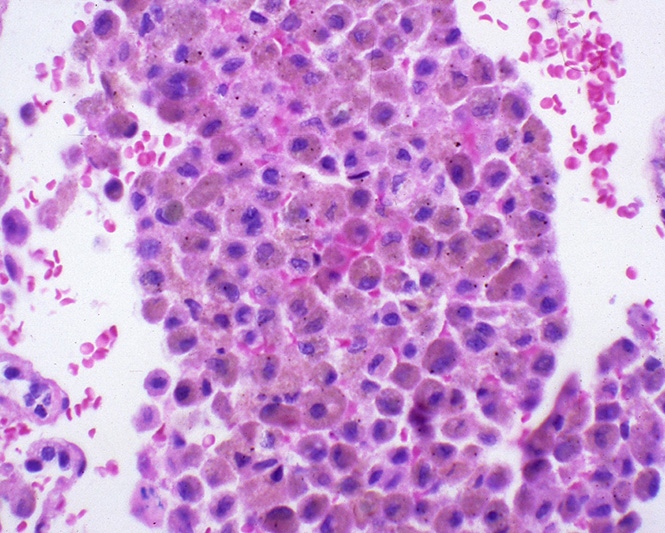
(Summary of Microscopic Findings - click here)
The tumor tissue consists of glandular structures with very large nuclei which are often arranged in several layers. The nuclear to cytoplasmic ratio is low. There are several mitoses and the stroma contains increased amounts of collagen. Well differentiated adenocarcinomas, with a glandular pattern like this one, are uncommon in the lung.
|
(Review Normal Histology - click here)
Norm No. 24 Lung
[ImageScope] [WebScope]
The primary function of the lung is gas exchange. Therefore, alveoli have thin walls lined by thin flat pneumocytes and endothelial cells. There is no thickening or fibrosis of the interstitium. The bronchioli are lined with basally oriented ciliated columnar epithelium. The bronchi are lined by similar epithelium. There are mucous glands within the submucosa. The bronchial smooth muscle is not hypertrophied. The pulmonary vessels are patent with no evidence of intimal thickening or muscular hyperplasia.
|
126-1. Which of the following is the most likely diagnosis?
- Adenocarcinoma
- Carcinoid tumor
- Large cell neuroendocrine carcinoma
- Small cell carcinoma
- Squamous cell carcinoma
ANSWER
126-2. Which of the following is true regarding this tumor?
- It arises from neuroendocrine cells
- It is the most common malignancy in non-smokers
- It is typically hilar
- Its incidence has decreased markedly in the last 20 years
- Mutations in N-RAS are common
ANSWER
CASE NUMBER 511
(no virtual slides for this case)
Clinical History: A 67-year-old man presented to his primary care physician with a two-month history of fatigue and weight loss of ten pounds. Clinical history was relevant for a 60-pack-year smoking history and a 5-year history of hypertension and obstructive pulmonary disease. A chest x-ray revealed a right hilar mass and a subsequent CT scan showed this to be a 10 cm mass with associated lymphadenopathy. A biopsy was performed.
Image Gallery:
511-1. Which of the following is the most likely diagnosis?
- Adenocarcinoma
- Carcinoid tumor
- Pulmonary hamartoma
- Small cell carcinoma
- Squamous cell carcinoma
ANSWER
511-2. Which of the following is true regarding this disease?
- Biologic behavior is typically indolent
- Cells show a neuroendocrine phenotype
- Hypercalcemia is commonly diagnosed in these patients
- Incidence among non-smokers is rising
- Surgical resection is standard therapy
ANSWER
511-3. Which of the following is most commonly associated with this disease?
- Carcinoid syndrome
- Horner syndrome
- Hypoglycemia
- Nephrotic syndrome
- Syndrome of inappropriate antidiuretic hormone secretion
ANSWER
511-4. Which of the following has the strongest association with cigarette smoking?
- Adenocarcinoma
- Adenosquamous carcinoma
- Pulmonary hamartoma
- Small cell carcinoma
- Squamous cell carcinoma
ANSWER
LUNG PATHOLOGY Review Items
Key Vocabulary Terms (click here to search any additional terms on Stedman's Online Medical Dictionary)
LEARNING OBJECTIVES
- Absolutely critical information you must know to practice medicine is in bold font.
- Important information that will be needed for routine patient care is in regular font.
- Information about less common diseases that you may encounter in clinical practice and that will probably appear on examinations is in italics
- Describe the mechanisms by which the following pulmonary defense mechanisms accomplish their functions:
- nasal clearance
- laryngeal (including epiglottic) action
- tracheobronchial clearance
- alveolar clearance
- Explain the pathogenesis of each of the manifestations of pulmonary disease:
|
- hypertrophic pulmonary osteoarthropathy
|
|
|
|
|
|
|
|
|
- Discuss the following pulmonary congenital anomalies, in terms of morphology and clinical consequences:
- agenesis
- hypoplasia
- congenital lobar overinflation ("emphysema")
- congenital cyst
- bronchopulmonary sequestration
- Contrast obstructive and restrictive pulmonary disease, in terms of:
- morphologic features
- radiologic manifestations
- pulmonary function test results
- clinical manifestations
- Compare and contrast the etiologies and effects of airflow obstruction that occur in lesions involving the airways with those that involve the alveolar parenchyma.
- Compare and contrast the clinical and pathologic features of:
- emphysema
- chronic bronchitis
- bronchial asthma
- bronchiectasis
- Compare and contrast the clinical and pathologic features of bronchial asthma:
- atopic
- non-atopic
- drug-induced
- occupational
- Compare and contrast the clinical and pathologic features of
:
- centriacinar (centrolobular) emphysema
|
|
- congenital lobar "emphysema"
|
|
- panacinar (panlobular) emphysema
|
|
- paraseptal (distal acinar) emphysema
|
|
- Discuss the Reid index.
- Discuss respiratory bronchiolitis of smokers (small airways disease) in terms of:
- pathogenesis
- morphology
- clinical presentation
- Discuss bronchiectasis, in terms of:
- predisposing conditions
- the types of organisms typically cultured from bronchi
- sequelae
- Compare and contrast neonatal and adult respiratory distress syndrome in terms of:
- predisposing factors/associated conditions
- pathogenesis
- morphology
- complications
- clinical course
- Compare and contrast the clinical and pathologic features of the following lung diseases:
- diffuse alveolar damage (DAD)
- bronchilitis obliterans-organizing pneumonia (BOOP)
- usual interstitial pneumonia (UIP)
- desquamative interstitial pneumonia (DIP)
- lymphoid interstitial pneumonia (LIP)
- nonspecific interstitial pneumonia (NSIP)
- Discuss the clinical and pathologic features of the following disorders:
- sarcoidosis
- Goodpasture syndrome
- idiopathic pulmonary hemosiderosis (IPH)
- hypersensitivity pneumonitis (HP)
- pulmonary alveolar proteinosis
- pulmonary eosinophilic granuloma
- pulmonary infiltrates with eosinophilia (PIE)
- lipid pneumonia
- Wegener granulomatosis
- lymphomatoid granulomatosis
- Discuss pulmonary invovement in autoimmune ("collagen-vascular") diseases, noting the major morphologic manifestations in the lung of:
- systemic lupus erythematosus (SLE)
- rheumatoid arthritis (RA)
- progessive systemic sclerosis (PSS)
- Discuss the basic pathogenesis of pneumoconioses.
- Compare and contrast the clinical and pathologic features of the following pneumoconioses:
- coal workers' pneumoconioses
- silicosis
- asbestosis
- berylliosis
- Discuss the clinical and pathologic features of these asbestos-related lung diseases:
- fibrous pleural plaques
- pleural effusion
- asbestosis
- bronchogenic carcinoma
- malignant mesothelioma
- Discuss the acute and chronic stages of radiation lung injury, in terms of:
- temporal features
- pathogenesis
- morphology
- consequences
- Discuss drug-induced lung disease, enumerating drugs most commonly associated with the following pulmonary reactions
- bronchospasm
- pulmonary edema
- hypersensitivity pneumonitis (HP)
- eosinophilic pneumonia
- diffuse alveolar damage (DAD)
- pulmonary fibrosis
- Enumerate the general indications for lung transplantation, and complications:
- pulmonary infection
- acute rejection
- chronic rejection
- Discuss the pulmonary features of cystic fibrosis (CF), in terms of:
- frequency of involvement of lung in CF
- pathogenesis
- morphology
- functional alterations
- clinical manifestations
- pulmonary complications
- obstructive
- infectious (including most common organisms involved)
- treatment
- prognosis
- Compare and contrast the clinical and pathologic features of:
- bronchopneumonia
- lobar pneumonia
- primary atypical pneumonia
- aspiration pneumonia
- lung abscess
- pulmonary infiltrates in the immunocompromised host
- Describe the four classic stages of the inflammatory response in lobar pneumonia in terms of:
- temporal features
- morphology
- Compare and contrast the clinical and pathologic features of :
|
|
|
|
|
|
|
- respiratory syncytial virus (RSV)
|
|
|
|
|
|
|
|
- acute respiratory syndrome
|
|
- Pneumocystis carinii pneumonia (PCP)
|
|
|
|
|
- Differentiate among tuberculosis, sarcoidosis, and granulomatous fungal disease
- etiopathogenesis
- morphologic features, including use of special stains
- organs involved
- radiologic features
- clinical presentation
- diagnostic tests
- laboratory findings
- prognosis
- Discuss pulmonary edema, embolism, and infarction in terms of:
- predisposing factors and etiology
- pathogenesis
- morphologic features
- radiologic features
- clinical manifestations
- Compare and contrast the clinical and pathologic features of pulmonary embolism:
- thrombus
- fat
- air
- bone marrow
- amniotic fluid
- talc
- Compare and contrast primary and secondary pulmonary hypertension, in terms of:
- predisposing factors/associated conditions
- pathogenesis
- age and sex distribution
- clinical manifestations
- size and type of vessels involved
- morphologic features (including reversible vs. irreversible lesions)
- hemodynamic consequences
- prognosis
- Discuss the clinical and pathologic features of:
- pulmonary circulatory disease associated with congenital heart disease
- persistent fetal circulation
- Compare and contrast the clinical and pathologic features of the following thoracic tumors:
- squamous cell carcinoma of lung
- bronchogeneic adenocarcinoma
- bronchioloalveolar carcinoma
- small cell carcinoma of lung
- large cell carcinoma of lung
- bronchial carcinoid
- pulmonary hamartoma
- malignant lymphoma
- Hodgkin disease
- metastatic neoplasm to thorax
- pleural fibroma (solitary fibrous tumor)
- malignant mesothelioma
- List likely etiologies and expected effects on pulmonary function of:
- hydrothorax
- empyema
- hemothorax
- chylothorax
- pneumothorax
- tension pneumothorax
- pleural adhesion
- Discuss pleural fluid collections on the basis of fluid type and common association.
- List appropriate diagnostic procedures for patients with pleural effusions.
- Compare and contrast the clinical and pathologic features of:
- nasal polyp
- sinonasal papilloma
- laryngeal nodule (singers' node)
- laryngeal papilloma
- juvenile laryngeal papillomatosis
- laryngeal squamous cell carcinoma
- nasopharyngeal carcinoma
|