CASE NUMBER 607
(no virtual slides for this case)
Clinical History: A 45-year old man comes to the emergency department in obvious severe pain. He states that he has severe pain in his left flank that extends down to his left groin. The pain is sharp and severe, and it started several minutes earlier.
607-1. What tests would you order?
- MRI of the flank
- Abdominal xray
- Intravenous pyelogram
- EKG
- Chest xray
ANSWER
Clinical History (continued): An abdominal radiograph is obtained as shown below.
Image Gallery:
607-2. What is the best diagnosis?
- Renal carcinoma
- Adrenal hemorrhage
- Foreign body / penetrating trauma
- Left ureteral urolithiasis
ANSWER
607-3. Which of the following is the most likely composition of the radiographic feature indicated by the arrow on the radiograph?
- Ammonium magnesium phosphate
- Calcium
- Cystine
- Uric acid
ANSWER
CASE NUMBER 202
[ImageScope] [WebScope]
Clinical History: A 74-year-old white man presented to his primary care physician with a one-year history of hesitancy, intermittency, nocturia and increasing difficulty in urination. Rectal examination revealed an enlarged, nodular firm prostate. A week after his visit, he experienced a myocardial infarction and died.
Image Gallery:
(Summary of Gross Findings - click here)
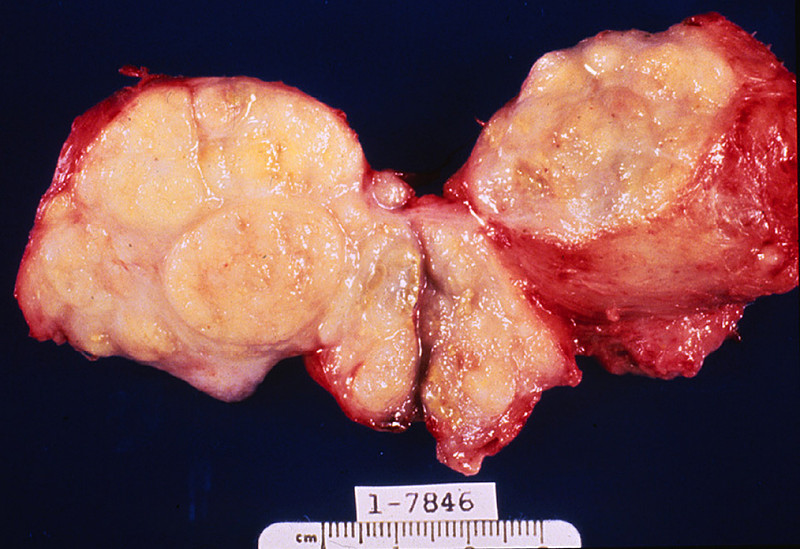
The prostate was large, nodular, and 120 grams in weight. The capsule was tense, and many gray-white firm nodules bulged out of the cut surface. These nodules varied from 3 mm to 1 cm. The larger ones were present in the lateral lobes and compressed the urethra.
|
(Summary of Microscopic Findings - click here)
Two types of nodules can be distinguished. One is composed essentially of fibro-muscular elements - stromal hyperplasia. The other is composed predominantly of epithelial glands. The size of the glands varies remarkably, and cystic dilatation of some is present. There are two types of cells forming the glands. The luminal cells are tall columnar cells with basal nuclei and apocrine secretory activity in the cytoplasm. The basal cell layer is composed of cuboidal or flattened epithelium. These glandular nodules are well demarcated by the encircling fibro-muscular stroma.
|
(Review Normal Histology - click here)
Norm No. 1 Prostate
[ImageScope] [WebScope]
The prostate gland is composed of multiple glandular spaces lines by a basal layer of cuboidal epithelium covered by columnar secretory cells with frequent papillary projections into the gland lumen. The cells are uniform in size and shape. The nuclei are not crowded. They do not have nucleoli that are visible. Glands are separated by fibrovascular stroma.
|
202-1. Which of the following is the most likely diagnosis?
- Adenocarcinoma of the prostate
- Benign prostatic hyperplasia
- Chronic prostatitis
- Foamy gland adenocarcinoma
- Metastatic clear cell renal carcinoma
ANSWER
202-2. Which of the following factors contributes most significantly to the development of this condition?
- Arylamine exposure
- Bladder stones
- Cigarette smoking
- Circulating androgens
- Hypertension
ANSWER
202-3. Patients with this condition are at significantly increased risk for which of the following?
- Adenocarcinoma of the prostate
- Condyloma acuminatum
- Priapism
- Pyelonephritis
- Spermatocytic seminoma
ANSWER
CASE NUMBER 5
[ImageScope] [WebScope]
Clinical History: A 77-year-old male went to his primary care physician for an annual checkup. Digital rectal exam revealed a firm nodule at the base of the prostate gland. Serum PSA was measured at 7.0 ng/mL (normal < 4.0 ng/mL). Needle biopsies were performed and the patient subsequently underwent a total prostatectomy.
Image Gallery:
(Summary of Gross Findings - click here)
The prostate was quite large and firm with multiple rubbery nodules measuring 2 mm to 6 mm in diameter. Some of the nodules contain yellowish flecks. The seminal vesicles were firm bilaterally.
|
(Summary of Microscopic Findings - click here)
There are a number of glands shown in varied patterns. In some cases the epithelial cells are found in non-glandular masses. The epithelial cells are cuboidal or polygonal with central, round, deeply pigmented nuclei. Few, if any, mitotic figures can be seen. The presence of perineural and perivascular invasion is clearly in evidence. This is a useful diagnostic characteristic of adenocarcinoma of the prostate.
|
(Review Normal Histology - click here)
Norm No. 1 Prostate
[ImageScope] [WebScope]
The prostate gland is composed of multiple glandular spaces lines by a basal layer of cuboidal epithelium covered by columnar secretory cells with frequent papillary projections into the gland lumen. The cells are uniform in size and shape. The nuclei are not crowded. They do not have nucleoli that are visible. Glands are separated by fibrovascular stroma.
|
5-1. Which of the following is the most likely diagnosis?
- Adenocarcinoma of the prostate
- Benign prostatic hypertrophy
- Chronic prostatitis
- Prostatic intraepithelial neoplasia (PIN)
- Small cell neuroendocrine carcinoma
ANSWER
5-2. Following his prostatectomy, the patient’s PSA level dropped to essentially zero. However, two years later, his serum PSA was again elevated. Which of the following is true concerning serum PSA?
- Current treatment recommendations include routine screening of PSA levels for all men over 65
- Even mildly elevated levels of PSA are highly suggestive of malignancy
- Following prostatectomy, serum PSA is a good marker of residual, recurrent or metastatic disease
- Serum PSA is a good screening test for undiagnosed prostate cancer
- Serum PSA is rarely elevated in benign conditions
ANSWER
5-3. Which of the following features is most characteristic of this lesion?
- Absence of a basal cell layer
- Extensive calcification
- Marked nuclear pleomorphism
- “Salt and pepper” chromatin
- Squamous pearls
ANSWER
5-4. Which of the following patients is at greatest risk for adenocarcinoma of the prostate?
- A 45-year-old Caucasian living in Ireland
- A 53-year-old Chinese living in Malaysia
- A 55-year-old Japanese living in the USA
- A 59-year-old African-American living in the USA
- A 60-year-old Thai living in Thailand
ANSWER
5-5. The Gleason grading system is based on which of the following features?
- Degree of gland formation
- Nuclear pleomorphism
- Number of mitotic figures
- Perineural invasion
- Presence of necrosis
ANSWER
5-6. Which of the following patients would benefit most from radical prostatectomy?
- 60 years old, Gleason score 6, stage T1b
- 60 years old, Gleason score 10, stage T2b
- 70 years old, Gleason score 6, stage T1b
- 70 years old, Gleason score 6, stage T3a
- 70 years old, Gleason score 10, stage T2b
ANSWER
CASE NUMBER 13
(no virtual slides for this case)
Clinical History: A 57-year-old man presented to his primary care physician after an episode of painless hematuria. Cystoscopy was performed and a mass was noted near the dome of the bladder.
Image Gallery:
13-1. Which of the following is the most likely diagnosis?
- Adenocarcinoma
- Papilloma
- Papillary urothelial carcinoma
- Small cell carcinoma
- Squamous cell carcinoma
ANSWER
13-2. Which of the following is the most significant risk factor in the development of carcinoma of the bladder?
- Arylamine exposure
- Bladder stones
- Cigarette smoking
- Schistosomiasis
- VHL mutation
ANSWER
13-3. A 35-year-old woman from Angola presents to her primary care physician following an episode of painless hematuria. A routine blood test revealed eosinophilia and subsequent ELISA analysis confirmed a diagnosis of schistosomiasis. This patient is at greatest risk for developing which of the following?
- Adenocarcinoma
- Papilloma
- Papillary urothelial carcinoma
- Small cell carcinoma
- Squamous cell carcinoma
ANSWER
CASE NUMBER 34
[ImageScope] [WebScope]
Clinical History: A 35-year-old Caucasian man presented to his primary care physician with non-specific testicular discomfort and stated that he has noticed that his right testicle felt larger than his left testicle. Physical exam revealed enlargement of the right testis, which did not transilluminate. The patient underwent a right orchiectomy.
Image Gallery:
(Summary of Gross Findings - click here)
The orchiectomy specimen has been opened to demonstate the soft fleshy tumor which has replaced the normal testis. The spermatic cord is attached.
|
(Summary of Microscopic Findings - click here)
Note the characteristic arrangement of cells in small groups outlined by delicate fibrous stroma. The cells are relatively uniform with distinct borders. The cytoplasm is clear and sometimes almost foamy. The cells are polygonal with large, round, central nuclei and prominent nucleoli. In many sections a fragment of atrophic and fibrotic testis may be seen adjacent to the tumor.
|
(Review Normal Histology - click here)
Norm No. 22 Testis
[ImageScope] [WebScope]
The testis is the male gland responsible for sperm production. There is a fibrous capsule and multiple glands separated by a loose stoma. Within the glands there are mature and immature sperm. Within the stroma there are a few large Leydig cells which are responsible for testosterone production.
|
34-1. Which of the following is the most likely diagnosis?
- Choriocarcinoma
- Embryonal carcinoma
- Leydig cell tumor
- Seminoma
- Yolk sac tumor
ANSWER
34-2. Elevated serum HCG levels in this patient would suggest which of the following?
- AFP will also be elevated
- More than one germ layer is present
- Radiation is the best therapy
- Schiller-Duval bodies will be seen microscopically
- This is a mixed tumor
ANSWER
34-3. Which of the following tumors is most sensitive to radiation therapy?
- Choriocarcinoma
- Embryonal carcinoma
- Sertoli cell tumor
- Seminoma
- Yolk sac tumor
ANSWER
34-4. An 18-year old Caucasian man presented to the emergency room with acute onset of left hemiplegia. Physical exam showed bilateral papilledema and an MRI revealed a right parietal lobar hematoma. Following evacuation of the hematoma, screening MRI revealed multiple lesions in the liver and spleen. In view of the possibility of metastatic disease, a careful physical exam was performed and found a 1 cm mass in the left testicle. Based on this clinical history, a biopsy of the testicular mass would most likely show which of the following?
- Choriocarcinoma
- Embryonal carcinoma
- Sertoli cell tumor
- Seminoma
- Yolk sac tumor
ANSWER
LOWER URINARY TRACT PATHOLOGY Review Items
Key Vocabulary Terms (click here to search any additional terms on Stedman's Online Medical Dictionary)
LEARNING OBJECTIVES
Absolutely critical information you must know to practice medicine is in bold font.
Important information that will be needed for routine patient care is in regular font.
Information about less common diseases that you may encounter in clinical practice and that will probably appear on examinations is in italics
- Describe the normal anatomy (gross and microscopic) of each of the following:
- Discuss the proper use of urinalysis in the evaluation of lower urinary tract disease, and interpret abnormalities of this test in clinical context
- Discuss obstruction at various levels of the urinary tract in terms of:
- site and nature of lesion
- etiology and pathogenesis
- alteration in renal function
- morphologic effect on kidney
- Discuss diverticula of the urinary bladder, in terms of:
- etiology
- pathogenesis
- morphology
- complications
- Discuss urolithiasis in terms of:
- relative incidence of various types of stones
- pathophysiologic abnormalities associated with the common types of stones
- etiology and pathogenesis of stone formation
- effect of location of stones on clinical and anatomic findings
- clinical course and complications
- Discuss the clinical and pathologic features of the following congenital anomalies:
- patent urachus
- hypospadias
- eductivepispadias
- exstrophy of the bladder
- duplications of the collecting system
- urethral valves
- Compare and contrast clinical and pathologic features of inflammatory conditions:
- infectious cystitis
- interstitial cystitis
- malacoplakia
- Compare and contrast the clinical and pathologic features of lower urinary tract neoplasms
- urothelial (transitional cell) carcinoma
- squamous cell carcinoma
- adenocarcinoma
MALE GENITAL SYSTEM Review Items
Key Vocabulary Terms (click here to search any additional terms on Stedman's Online Medical Dictionary)
LEARNING OBJECTIVES
Absolutely critical information you must know to practice medicine is in bold font.
Important information that will be needed for routine patient care is in regular font.
Information about less common diseases that you may encounter in clinical practice and that will probably appear on examinations is in italics
- Compare and contrast the following congenital anomalies:
- Discuss the clinical and pathologic features of the following neoplasms:
- squamous cell carcinoma of penis and scrotum
- adenocarcinoma of prostate
- germ cell tumors of testis
- sex cord-stromal tumors of testis
- malignant lymphoma of testis
- Compare and contrast the clinical and pathologic features of the following inflammatory conditions:
- prostatitis (acute, chronic granulomatous)
- orchitis (nonspecific, mumps, granulomatous)
- torsion of spermatic cord
- Discuss the clinical and pathologic features of the following disorders:
- nodular hyperplasia of the prostate
- cryptorchidism
- Classify anatomically the causes of male infertility.
|